HDAC1 抗体
 (7 references)
 (1 validation)
Our Local Distributor
北京 101111
Quick Overview for HDAC1 抗体 (ABIN2854776)
抗原
See all HDAC1 抗体适用
宿主
克隆类型
标记
应用范围
质量等级
-
-
交叉反应
- 人, 小鼠, 大鼠, 斑马鱼
-
产品特性
-
Rabbit Polyclonal antibody to HDAC1 (histone deacetylase 1)
HDAC1 antibody -
纯化方法
- Purified by antigen-affinity chromatography.
-
免疫原
- Recombinant protein encompassing a sequence within the center region of human HDAC1. The exact sequence is proprietary.
-
亚型
- IgG
-
-
anti-Histone Deacetylase 1 (HDAC1) (AA 1-482) antibody
HDAC1 适用: 人 WB, ELISA, IF, IHC (p), PLA 宿主: 小鼠 Monoclonal 5C11 unconjugated
anti-Histone Deacetylase 1 (HDAC1) (AA 1-482) antibodyHDAC1 适用: 人 WB, IHC, IF, ICC, FACS 宿主: 小鼠 Monoclonal E22 unconjugated
anti-Histone Deacetylase 1 (HDAC1) (AA 1-482) antibodyHDAC1 适用: 人 IHC, ELISA, IF, ChIP 宿主: 兔 Polyclonal unconjugated
anti-Histone Deacetylase 1 (HDAC1) antibodyKO Validated HDAC1 适用: 人 WB, IHC, IF 宿主: 兔 Monoclonal unconjugated
anti-Histone Deacetylase 1 (HDAC1) (AA 1-482) antibodyHDAC1 适用: 人 WB, ELISA, IF, IHC (p) 宿主: 小鼠 Monoclonal 5A11 unconjugated
anti-Histone Deacetylase 1 (HDAC1) (AA 450-482) antibodyHDAC1 适用: 人 WB, IHC, ELISA, IF, FM, MA 宿主: 兔 Polyclonal unconjugated
anti-Histone Deacetylase 1 (HDAC1) (C-Term) antibodyVerified HDAC1 适用: 人, 小鼠 WB, ELISA, IF, ChIP 宿主: 山羊 Polyclonal unconjugated
anti-Histone Deacetylase 1 (HDAC1) (Middle Region) antibodyHDAC1 适用: 人, 小鼠, 大鼠, 兔, Cow, 犬, 豚鼠, 马, 斑马鱼, Saccharomyces cerevisiae WB, IHC 宿主: 兔 Polyclonal unconjugated
anti-Histone Deacetylase 1 (HDAC1) (C-Term) antibodyHDAC1 适用: 人, 小鼠, 大鼠 WB, IHC, ELISA, IF, ICC 宿主: 兔 Polyclonal unconjugated
anti-Histone Deacetylase 1 (HDAC1) (AA 271-477) antibodyHDAC1 适用: 人, 小鼠 WB, IHC, IF, ICC, IHC (p) 宿主: 兔 Polyclonal unconjugated
-
-
应用备注
- WB: 1:500-1:3000. ICC/IF: 1:100-1:1000. IHC-P: 1:100-1:1000. IP: 1:100-1:500. Optimal dilutions/concentrations should be determined by the researcher. Not tested in other applications.
-
说明
-
Positive Control: 293T , A431 , HeLa , HepG2 , U87-MG , SK-N-SH , Rat-2 , NIH3T3 , DDDDK-tagged HDAC1-transfected 293T
Validation: KO/KD, Orthogonal, Overexpression
-
限制
- 仅限研究用
-
-
- by
- Gianluca Zambanini, Anna Nordin and Claudio Cantù; Cantù Lab, Gene Regulation during Development and Disease, Linköping University
- No.
- #104404
- 日期
- 2022.02.28
- 抗原
- HDAC1
- Lot Number
- Method validated
- Cleavage Under Targets and Release Using Nuclease
- Positive Control
Polyclonal rabbit anti-H3K4me (antibodies-online, ABIN3023251)
- Negative Control
Polyclonal guinea pig anti-rabbit IgG (antibodies-online, ABIN101961)
- Notes
Passed. ABIN2854776 allows for HDAC1 targeted digestion using CUT&RUN in mouse fore limbs (11.5) cells.
- Primary Antibody
- ABIN2854776
- Secondary Antibody
- Full Protocol
- Cell harvest and nuclear extraction
- Dissect 3 Fore limbs (11.5 DAC) from mouse strain RjOrl:SWISS for each sample.
- Dissociate the tissue into single cells in TrypLE for 15 min at 37 °C.
- Centrifuge cell solution 5 min at 800 x g at RT.
- Remove the liquid carefully.
- Gently resuspend cells in 1 mL of Nuclear Extraction Buffer (20 mM HEPES-KOH pH 8.2, 20% Glycerol, 0,05% IGEPAL, 0.5 mM Spermidine, 10 mM KCl, Roche Complete Protease Inhibitor EDTA-free).
- Move the solution to a 2 mL centrifuge tube.
- Pellet the nuclei 800 x g for 5 min.
- Repeat the NE wash twice for a total of three washes.
- Resuspend the nuclei in 20 µL NE Buffer per sample.
- Concanavalin A beads preparation
- Prepare one 2 mL microcentrifuge tube.
- Gently resuspend the magnetic Concanavalin A Beads (antibodies-online, ABIN6952467).
- Pipette 20 µL Con A Beads slurry for each sample into the 2 mL microcentrifuge tube.
- Place the tube on a magnet stand until the fluid is clear. Remove the liquid carefully.
- Remove the microcentrifuge tube from the magnetic stand.
- Pipette 1 mL Binding Buffer (20 mM HEPES pH 7.5, 10 mM KCl, 1 mM CaCl2, 1 mM MnCl2) into the tube and resuspend ConA beads by gentle pipetting.
- Spin down the liquid from the lid with a quick pulse in a table-top centrifuge.
- Place the tubes on a magnet stand until the fluid is clear. Remove the liquid carefully.
- Remove the microcentrifuge tube from the magnetic stand.
- Repeat the wash twice for a total of three washes.
- Gently resuspend the ConA Beads in a volume of Binding Buffer corresponding to the original volume of bead slurry, i.e. 20 µL per sample.
- Nuclei immobilization – binding to Concanavalin A beads
- Carefully vortex the nuclei suspension and add 20 µL of the Con A beads in Binding Buffer to the cell suspension for each sample.
- Close tube tightly incubates 10 min at 4 °C.
- Put the 2 mL tube on the magnet stand and when the liquid is clear remove the supernatant.
- Resuspend the beads in 1 mL of EDTA Wash buffer (20 mM HEPES pH 7.5, 150 mM NaCl, 0.5 mM Spermidine, Roche Complete Protease Inhibitor EDTA-free, 2mM EDTA).
- Incubate 5 min at RT.
- Place the tube on the magnet stand and when the liquid is clear remove the supernatant.
- Resuspend the beads in 200 µl of Wash Buffer (20 mM HEPES pH 7.5, 150 mM NaCl, 0.5 mM Spermidine, Roche Complete Protease Inhibitor EDTA-free) per sample.
- Primary antibody binding
- Divide nuclei suspension into separate 200 µL PCR tubes, one for each antibody.
- Add 2 µL antibody (anti-HDAC1 antibody ABIN2854776, anti-H3K27me3 antibody positive control ABIN6923144, and guinea pig anti-rabbit IgG negative control antibody ABIN101961) to the respective tube, corresponding to a 1:100 dilution.
- Incubate at 4 °C ON.
- Place the tubes on a magnet stand until the fluid is clear. Remove the liquid carefully.
- Remove the microcentrifuge tubes from the magnetic stand.
- Wash with 200 µL of Wash Buffer using a multichannel pipette to accelerate the process.
- Repeat the wash five times for a total of six washes.
- pAG-MNase Binding
- Prepare a 1.5 mL microcentrifuge tube containing 100 µL of pAG mix per sample (100 µL of wash buffer + 58.5 µg pAG-MNase per sample).
- Place the PCR tubes with the sample on a magnet stand until the fluid is clear. Remove the liquid carefully.
- Remove tubes from the magnetic stand.
- Resuspend the beads in 100 µL of pAG-MNase premix.
- Incubate 30 min at 4 °C.
- Place the tubes on a magnet stand until the fluid is clear. Remove the liquid carefully.
- Remove the microcentrifuge tubes from the magnetic stand.
- Wash with 200 µL of Wash Buffer using a multichannel pipette to accelerate the process.
- Repeat the wash five times for a total of six washes.
- Resuspend in 100 µL of Wash Buffer.
- MNase digestion and release of pAG-MNase-antibody-chromatin complexes
- Place PCR tubes on ice and allow to chill.
- Prepare a 1.5 mL microcentrifuge tube with 102 µl of 2 mM CaCl2 mix per sample (100 µl Wash Buffer + 2 µL 100 mM CaCl2) and let it chill on ice.
- Always in ice, place the samples on the magnetic rack and when the liquid is clear remove the supernatant.
- Resuspend the samples in 100 µl of the 2 mM CaCl2 mix and incubate in ice for exactly 30 min.
- Place the sample on the magnet stand and when the liquid is clear remove the supernatant.
- Resuspend the sample in 50 µl of 1x Urea STOP Buffer (8.5 M Urea, 100 mM NaCl, 2 mM EGTA, 2 mM EDTA, 0,5% IGEPAL).
- Incubate the samples 1h at 4°C.
- Transfer the supernatant containing the pAG-MNase-bound digested chromatin fragments to fresh 200 µl PCR tubes.
- DNA Clean up
- Take the Mag-Bind® TotalPure NGS beads (Omega Bio-Tek, M1378-01) from the storage and wait until they are at RT.
- Add 2x volume of beads to each sample (e.g. 100 µL of beads for 50 µL of sample).
- Incubate the beads and the sample for 15 min at RT.
- During incubation prepare fresh EtOH 80%.
- Place the PCR tubes on a magnet stand and when the liquid is clear remove the supernatant.
- Add 200 µl of fresh 80% EtOH to the sample without disturbing the beads (Important!!! Do NOT resuspend the beads or remove the tubes from the magnet stand or the sample will be lost).
- Incubate 30 sec at RT.
- Remove the EtOH from the sample.
- Repeat the wash with 80% EtOH.
- Resuspend the beads in 25 µL of 10 mM Tris.
- Incubate the sample for 2 min at RT.
- Repeat the 2x beads clean up as described before (this time with 50 µL of beads for each sample).
- Resuspend the beads + DNA in 20 µL of 10 mM Tris.
- Library preparation and sequencing
- Prepare Libraries using KAPA HyperPrep Kit using KAPA Dual-Indexed adapters according to protocol.
- Sequence samples on an Illumina NextSeq 500 sequencer, using a NextSeq 500/550 High Output Kit v2.5 (75 Cycles), 36 bp PE.
- Peak calling
- Trim reads using using bbTools bbduk (BBMap - Bushnell B. - sourceforge.net/projects/bbmap/) to remove adapters, artifacts and repeat sequences.
- Map aligned reads to the hg38 human genome using bowtie with options -m 1 -v 0 -I 0 -X 500.
- Use SAMtools to convert SAM files to BAM files and remove duplicates.
- Use BEDtools genomecov to produce Bedgraph files.
- Call peaks using SEACR with a 0.001 threshold and the option norm stringent.
- Experimental Notes
The protocol is published in Zambanini, G. et al. A New CUT&RUN Low Volume-Urea (LoV-U) protocol uncovers Wnt/β-catenin tissue-specific genomic targets. bioRxiv (2022). https://doi.org/10.1101/2022.07.06.498999
生效 #104404 (Cleavage Under Targets and Release Using Nuclease)
 Validation Images
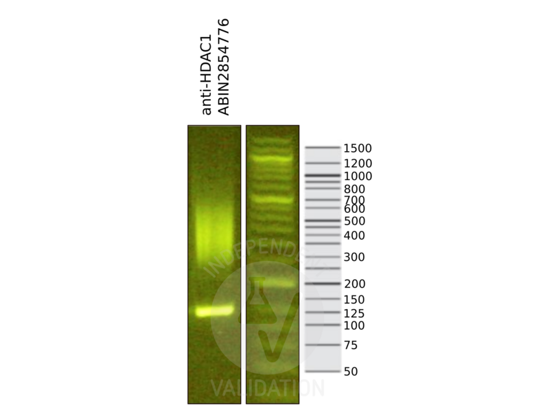
![Library profiles comparing fragment size distributions on an E-Gel EX 2% agarose gel (Thermo Fisher). Fragments obtained from CUT&RUN using anti-HDAC1 antibody ABIN2854776 after library preparation, compared to the E-Gel Sizing DNA Ladder (Thermo Fisher).]() Library profiles comparing fragment size distributions on an E-Gel EX 2% agarose gel (Thermo Fisher). Fragments obtained from CUT&RUN using anti-HDAC1 antibody ABIN2854776 after library preparation, compared to the E-Gel Sizing DNA Ladder (Thermo Fisher).
Library profiles comparing fragment size distributions on an E-Gel EX 2% agarose gel (Thermo Fisher). Fragments obtained from CUT&RUN using anti-HDAC1 antibody ABIN2854776 after library preparation, compared to the E-Gel Sizing DNA Ladder (Thermo Fisher).
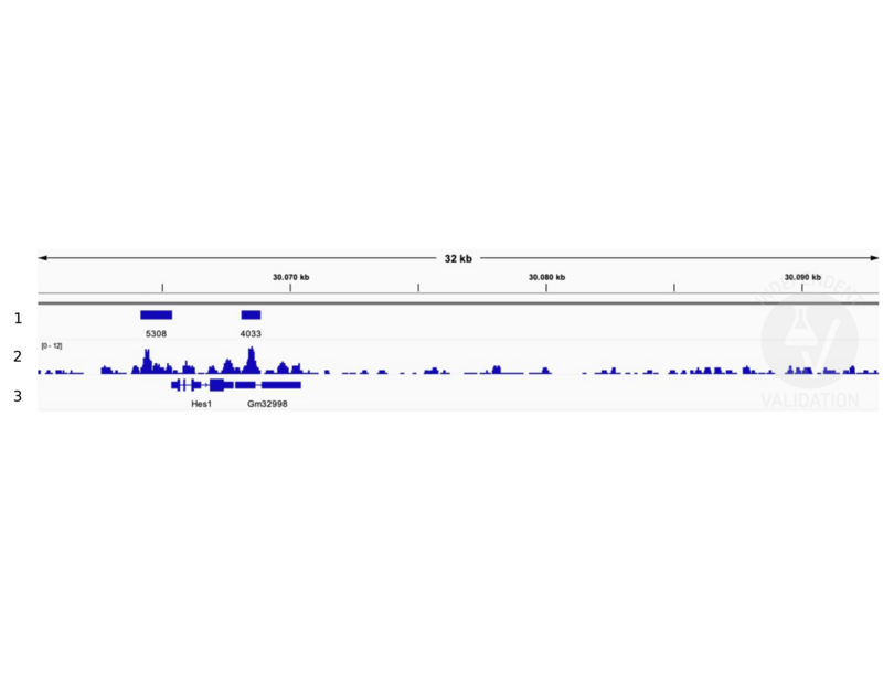
![1. Alignment tracks from CUT&RUN targeting HDAC1 in Mouse fore limb (11.5) cells using anti-HDAC1 antibody ABIN2854776. 2. Peaks called by SEACR from CUT&RUN data using anti HDAC1 ABIN2854776. 3. RefSeq Genes.]() 1. Alignment tracks from CUT&RUN targeting HDAC1 in Mouse fore limb (11.5) cells using anti-HDAC1 antibody ABIN2854776. 2. Peaks called by SEACR from CUT&RUN data using anti HDAC1 ABIN2854776. 3. RefSeq Genes.
Full Methods
1. Alignment tracks from CUT&RUN targeting HDAC1 in Mouse fore limb (11.5) cells using anti-HDAC1 antibody ABIN2854776. 2. Peaks called by SEACR from CUT&RUN data using anti HDAC1 ABIN2854776. 3. RefSeq Genes.
Full Methods -
-
状态
- Liquid
-
浓度
- 1 mg/mL
-
缓冲液
- 1XPBS ( pH 7), 20 % Glycerol, 0.01 % Thimerosal
-
储存液
- Thimerosal (Merthiolate)
-
注意事项
- This product contains Thimerosal (Merthiolate): a POISONOUS AND HAZARDOUS SUBSTANCE which should be handled by trained staff only.
-
储存条件
- 4 °C,-20 °C
-
储存方法
- Store as concentrated solution. Centrifuge briefly prior to opening vial. For short-term storage (1-2 weeks), store at 4°C. For long-term storage, aliquot and store at -20°C or below. Avoid multiple freeze-thaw cycles.
-
-
-
: "A new cut&run low volume-urea (LoV-U) protocol optimized for transcriptional co-factors uncovers Wnt/b-catenin tissue-specific genomic targets." in: Development (Cambridge, England), (2022) (PubMed).
: "Differential Expression of Multiple Disease-Related Protein Groups Induced by Valproic Acid in Human SH-SY5Y Neuroblastoma Cells." in: Brain sciences, Vol. 10, Issue 8, (2020) (PubMed).
: "Genome-wide kinetic properties of transcriptional bursting in mouse embryonic stem cells." in: Science advances, Vol. 6, Issue 25, pp. eaaz6699, (2020) (PubMed).
: "Nifedipine Exacerbates Lipogenesis in the Kidney via KIM-1, CD36, and SREBP Upregulation: Implications from an Animal Model for Human Study." in: International journal of molecular sciences, Vol. 21, Issue 12, (2020) (PubMed).
: "Overexpression of peptidase inhibitor 16 attenuates angiotensin II-induced cardiac fibrosis via regulating HDAC1 of cardiac fibroblasts." in: Journal of cellular and molecular medicine, Vol. 24, Issue 9, pp. 5249-5259, (2020) (PubMed).
: "The chromatin remodeler RSF1 controls centromeric histone modifications to coordinate chromosome segregation." in: Nature communications, Vol. 9, Issue 1, pp. 3848, (2019) (PubMed).
: "Inhibition of HDAC3- and HDAC6-Promoted Survivin Expression Plays an Important Role in SAHA-Induced Autophagy and Viability Reduction in Breast Cancer Cells." in: Frontiers in pharmacology, Vol. 7, pp. 81, (2016) (PubMed).
-
: "A new cut&run low volume-urea (LoV-U) protocol optimized for transcriptional co-factors uncovers Wnt/b-catenin tissue-specific genomic targets." in: Development (Cambridge, England), (2022) (PubMed).
-
- HDAC1 (Histone Deacetylase 1 (HDAC1))
-
别名
- histone deacetylase 1
-
背景
-
Histone acetylation and deacetylation, catalyzed by multisubunit complexes, play a key role in the regulation of eukaryotic gene expression. The protein encoded by this gene belongs to the histone deacetylase/acuc/apha family and is a component of the histone deacetylase complex. It also interacts with retinoblastoma tumor-suppressor protein and this complex is a key element in the control of cell proliferation and differentiation. Together with metastasis-associated protein-2, it deacetylates p53 and modulates its effect on cell growth and apoptosis.
Cellular Localization: Nucleus -
分子量
- 55 kDa
-
基因ID
- 3065
-
UniProt
- Q13547
-
途径
- Neurotrophin Signaling Pathway, Intracellular Steroid Hormone Receptor Signaling Pathway, Regulation of Intracellular Steroid Hormone Receptor Signaling, Mitotic G1-G1/S Phases, Regulation of Muscle Cell Differentiation, Skeletal Muscle Fiber Development, Negative Regulation of intrinsic apoptotic Signaling, Embryonic Body Morphogenesis
抗原
-


