CDKN1B 抗体


 (2 validations)
Our Local Distributor
北京 101111
Quick Overview for CDKN1B 抗体 (ABIN3025539)
抗原
See all CDKN1B 抗体适用
宿主
克隆类型
标记
应用范围
克隆位点
-
-
原理
- p27 Antibody Cocktail
-
纯化方法
- Protein G affinity chromatography
-
免疫原
- Mouse recombinant protein (DCS-72.F6) and recombinant human protein (KIP1/769) were used as the immunogen for the p27 antibody cocktail.
-
亚型
- IgG1, kappa
-
-
anti-Cyclin-Dependent Kinase Inhibitor 1B (p27, Kip1) (CDKN1B) (AA 150-198) antibody
CDKN1B 适用: 人, 小鼠, 大鼠 WB, ELISA, FACS, IHC (p), IF (cc), IF (p), IHC (fro) 宿主: 兔 Polyclonal unconjugated
anti-Cyclin-Dependent Kinase Inhibitor 1B (p27, Kip1) (CDKN1B) (AA 1-198) antibodyCDKN1B 适用: 人 WB, ELISA, IF, IHC (p), PLA 宿主: 小鼠 Monoclonal 4B4-E6 unconjugated
anti-Cyclin-Dependent Kinase Inhibitor 1B (p27, Kip1) (CDKN1B) antibodyKO Validated CDKN1B 适用: 人 WB, IHC, IF, IHC (p), IP, ICC, IHC (fro) 宿主: 兔 Polyclonal unconjugated
anti-Cyclin-Dependent Kinase Inhibitor 1B (p27, Kip1) (CDKN1B) antibodyCDKN1B 适用: 人, 小鼠, 大鼠, 猴 WB, IHC, IF, FACS 宿主: 小鼠 Monoclonal SX53G8 unconjugated
anti-Cyclin-Dependent Kinase Inhibitor 1B (p27, Kip1) (CDKN1B) antibodyKD Validated CDKN1B 适用: 人 WB, FACS, ICC 宿主: 兔 Monoclonal 23GB1825 unconjugated Recombinant Antibody
anti-Cyclin-Dependent Kinase Inhibitor 1B (p27, Kip1) (CDKN1B) (AA 1-198) antibodyCDKN1B 适用: 人 WB, IF 宿主: 小鼠 Polyclonal unconjugated
anti-Cyclin-Dependent Kinase Inhibitor 1B (p27, Kip1) (CDKN1B) (pThr187) antibodyCDKN1B 适用: 人, 小鼠, 大鼠 WB, IHC, ELISA, IF, ICC 宿主: 兔 Polyclonal unconjugated
anti-Cyclin-Dependent Kinase Inhibitor 1B (p27, Kip1) (CDKN1B) antibodyCDKN1B 适用: 人, 小鼠, 大鼠, 猴 WB, IHC, IF, FACS 宿主: 小鼠 Monoclonal DCS-72-F6 unconjugated
anti-Cyclin-Dependent Kinase Inhibitor 1B (p27, Kip1) (CDKN1B) antibodyCDKN1B 适用: 人 WB, IHC, ELISA, IF 宿主: 兔 Monoclonal 1G2 unconjugated Recombinant Antibody
anti-Cyclin-Dependent Kinase Inhibitor 1B (p27, Kip1) (CDKN1B) (AA 1-198) antibodyCDKN1B 适用: 人 WB, IF 宿主: 兔 Polyclonal unconjugated
-
-
应用备注
-
Optimal dilution of the p27 antibody to be determined by the researcher.
1. Staining of formalin-fixed tissues requires boiling tissue sections in 10 mM Citrate buffer, pH 6.0, for 10-20 min followed by cooling at RT for 20 min
2. The prediluted format is supplied in a dropper bottle and is optimized for use in IHC. After epitope retrieval step (if required), drip mAb solution onto the tissue section and incubate at RT for 30 min. -
限制
- 仅限研究用
-
-
- by
- Johann-Friedrich-Blumenbach-Institute for Zoology and Anthropology, Department of Developmental Biology, Georg-August-University Göttingen
- No.
- #101901
- 日期
- 2017.12.04
- 抗原
- P27
- Lot Number
- V2438-171009
- Method validated
- Western Blotting
- Positive Control
- NIH/3T3 mouse embryonic fibroblast cells overexpressing mVenus-tagged p27K, starved for 48h
- Negative Control
- Untransfected NIH/3T3 cells, starved for 48h
- Notes
Passed. ABIN3025539 detects the ectopically expressed fusion protein as well as the endogenous protein by immunoblotting. Unspecific cross-reactivity is low.
- Primary Antibody
- ABIN3025539
- Secondary Antibody
- anti-mouse IgG (whole molecule), HRP-linked (Sigma-Aldrich, A9044, lot 034M4761)
- Full Protocol
- Grow NIH/3T3 cells (ATCC, CRL-1658) in DMEM+GlutaMAX (Gibco, 31966-021, Lot 1852045) supplemented with fetal bovine serum (Gibco 270-106) and Pen/Strep (Gibco 15140), at 37°C and 5% CO2 to 70% confluency.
- Transfect cells with a plasmid encoding mVenus-tagged p27K- (kindly provided by Toshio Kitamura, University of Tokyo; Oki et al., 2014) using EndofectinMax (GeneCopoeia) following the manufacturer's instructions.
- Serum starve cells for 48h. Use untransfected NIH/3T3 cells starved for 48h as control.
- Lyse cells in RIPA buffer (10mM PBS pH7.2, 2mM EDTA, 1% NP-40, 1% Triton X-100, protease inhibitors) at 4°C.
- Denature total cellular lysates proteins in 1x SDS-sample buffer and separate proteins on a freshly cast denaturing 10% SDS-PAGE (Laemmli, 1970).
- Transfer proteins onto 0.2µm Protran membrane (GE Healthcare, 10600004, A10043108) with a Western blotting system for 1h at 400A (Towbin et al., 1979).
- Block the membrane in TBST (50mM Tris-HCl, pH7.4, 150mM NaCl, 0.2% Tween 20) containing 5% milk (blocking solution) for 60min at RT.
- Incubate membrane with primary mouse anti-P27 antibody (antibodies-online, ABIN3025539, lot V2438-171009) diluted 1:250 in blocking solution ON at 4°C.
- Incubate membrane with secondary anti-mouse IgG (whole molecule), HRP-linked (Sigma-Aldrich, A9044, lot 034M4761) diluted 1:5000 in TBST for 45min at RT.
- Wash membrane with TBST for 30-45min at RT.
- Reveal protein bands using Clarity Max Western ECL substrate (Bio-Rad, 1705062); image capture via Chemidoc Imaging System (BioRad).
- Experimental Notes
ABIN3025539 reveals a protein band at the expected molecular weight of the mVenus-p27 fusion protein. The antibody does also detect a protein at the expected molecular weight of the endogenous, untagged p27.
生效 #101901 (Western Blotting)
 Validation Images
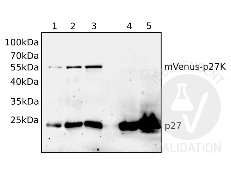
![Different volumes of total cellular lysates of either mVenus-p27K- transfected NIH/3T3 cells (1, 2, 3) or untransfected NIH/3T3 cells (4, 5) were loaded and the blot membrane was incubated with ABIN3025539 followed by incubation with anti-mouse IgG-linked to HRP and chemiluminescence detection. Expected molecular weight of p27 fused to mVenus is approximately 55kDa.]() Different volumes of total cellular lysates of either mVenus-p27K- transfected NIH/3T3 cells (1, 2, 3) or untransfected NIH/3T3 cells (4, 5) were loaded and the blot membrane was incubated with ABIN3025539 followed by incubation with anti-mouse IgG-linked to HRP and chemiluminescence detection. Expected molecular weight of p27 fused to mVenus is approximately 55kDa.
Full Methods
Different volumes of total cellular lysates of either mVenus-p27K- transfected NIH/3T3 cells (1, 2, 3) or untransfected NIH/3T3 cells (4, 5) were loaded and the blot membrane was incubated with ABIN3025539 followed by incubation with anti-mouse IgG-linked to HRP and chemiluminescence detection. Expected molecular weight of p27 fused to mVenus is approximately 55kDa.
Full Methods -
- by
- Johann-Friedrich-Blumenbach-Institute for Zoology and Anthropology, Department of Developmental Biology, Georg-August-University Göttingen
- No.
- #102346
- 日期
- 2017.12.04
- 抗原
- P27
- Lot Number
- V2438-171009
- Method validated
- Immunocytochemistry
- Positive Control
- NIH/3T3 mouse embryonic fibroblast cells overexpressing mVenus-tagged p27K, starved for 48h
- Negative Control
- Untransfected NIH/3T3 cells, starved for 48h
- Notes
Passed. ABIN3025539 detects the ectopically expressed fusion protein by immunocytochemistry. Unspecific cross-reactivity is low.
- Primary Antibody
- ABIN3025539
- Secondary Antibody
- goat anti-mouse IgG (H+L) Alexa Fluor 555 (Invitrogen, A21422, lot 948498)
- Full Protocol
- Grow NIH/3T3 cells (ATCC, CRL-1658) in on cover slips in DMEM, 10% fetal bovine serum (Gibco 270-106), 5% penicillin/streptomycin (Gibco) at 37°C in 5% CO2.
- Transfect cells with a plasmid encoding mVenus-tagged p27K- (kindly provided by Toshio Kitamura, University of Tokyo; Oki et al., 2014) using EndofectinMax (GeneCopoeia) following the manufacturer's instructions.
- Serum starve cells for 48h. Use untransfected NIH/3T3 cells starved for 48h as control.
- Fix cells in 3.7% paraformaldehyde (in PBS) for 15min at 4°C followed by incubation in 0.3% Triton X-100 for 10min.
- Block unspecific binding sites in PBT (phosphate buffered saline (PBS) containing 1% bovine serum albumin, 0.5% Tween-20) for 1h at RT.
- Incubate cells with primary mouse anti-P27 antibody (antibodies-online, ABIN3025539, lot V2438-171009) diluted 1:100 in PBS ON at 4°C.
- Wash cells with TBST (50mM Tris-HCl, pH7.4, 150mM NaCl, 0.1% Tween 20) for 15min.
- Incubate cells with secondary antibody goat anti-mouse IgG (H+L) Alexa Fluor 555 (Invitrogen, A21422, lot 948498) diluted 1:1000 in PBS and DAPI (4’,6-Diamidino-2-phenylindole; Sigma D-9542).
- Image acquisition on Zeiss LSM 510 confocal microscope and processing using Adobe Photoshop 5.0.
- Experimental Notes
生效 #102346 (Immunocytochemistry)
 Validation Images
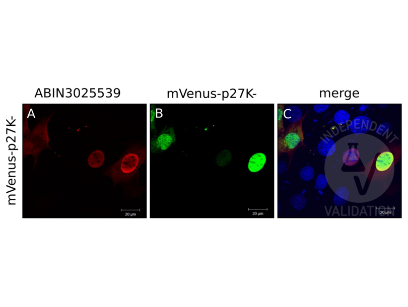
![NIH/3T3 cells were transfected with a plasmid encoding mVenus fused to p27K- and the expressed fusion protein detected by Venus autofluorescence (B, green) and by immunocytology using ABIN3025539 and anti-mouse AF555 (A, red). C shows red and green channels merged with DAPI counterstain (blue).]() NIH/3T3 cells were transfected with a plasmid encoding mVenus fused to p27K- and the expressed fusion protein detected by Venus autofluorescence (B, green) and by immunocytology using ABIN3025539 and anti-mouse AF555 (A, red). C shows red and green channels merged with DAPI counterstain (blue).
Full Methods
NIH/3T3 cells were transfected with a plasmid encoding mVenus fused to p27K- and the expressed fusion protein detected by Venus autofluorescence (B, green) and by immunocytology using ABIN3025539 and anti-mouse AF555 (A, red). C shows red and green channels merged with DAPI counterstain (blue).
Full Methods -
-
状态
- Liquid
-
浓度
- 0.2 mg/mL
-
缓冲液
- 0.2 mg/mL in 1X PBS with 0.1 mg/mL BSA (US sourced) and 0.05 % sodium azide
-
储存液
- Sodium azide
-
注意事项
- This product contains Sodium azide: a POISONOUS AND HAZARDOUS SUBSTANCE which should be handled by trained staff only.
-
储存条件
- 4 °C,-20 °C
-
储存方法
- Store the p27 antibody cocktail at 2-8oC (with azide) or aliquot and store at -20oC or colder (without azide).
-
-
- CDKN1B (Cyclin-Dependent Kinase Inhibitor 1B (p27, Kip1) (CDKN1B))
-
别名
- Cyclin-Dependent Kinase Inhibitor 1B (p27, Kip1)
-
背景
- Recognizes a 27 kDa protein, identified as the p27Kip1, a cell cycle regulatory mitotic inhibitor. It is highly specific and shows no cross-reaction with other related mitotic inhibitors. p27Kip1 functions as a negative regulator of G1 progression and has been proposed to function as a possible mediator of TGF- induced G1 arrest. p27Kip1 is a candidate tumor suppressor gene. This mAb co-precipitates cdk4 in complex p27Kip1 and is excellent for staining of formalin-fixed tissues.
-
UniProt
- P46527
-
途径
- Cell Division Cycle, Fc-epsilon Receptor Signaling Pathway, EGFR Signaling Pathway, Neurotrophin Signaling Pathway, Positive Regulation of Peptide Hormone Secretion, Negative Regulation of Hormone Secretion, Sensory Perception of Sound, Mitotic G1-G1/S Phases, DNA Replication, Positive Regulation of Endopeptidase Activity, Synthesis of DNA, Autophagy
抗原
-