HYAL1 抗体 (N-Term)
 (1 validation)
Our Local Distributor
北京 101111
Quick Overview for HYAL1 抗体 (N-Term) (ABIN2784342)
抗原
See all HYAL1 抗体适用
宿主
克隆类型
标记
应用范围
-
-
抗原表位
- N-Term
-
序列
- WNANTQWCLE RHGVDVDVSV FDVVANPGQT FRGPDMTIFY SSQLGTYPYY
-
预测反应
- Cow: 83%, Dog: 86%, Guinea Pig: 100%, Horse: 100%, Human: 100%, Mouse: 100%, Rabbit: 93%, Rat: 100%
-
产品特性
- This is a rabbit polyclonal antibody against HYAL1. It was validated on Western Blot using a cell lysate as a positive control.
-
纯化方法
- Affinity Purified
-
免疫原
- The immunogen is a synthetic peptide directed towards the N terminal region of human HYAL1
-
-
anti-Hyaluronidase-1 (HYAL1) (N-Term) antibody
HYAL1 适用: 人, 小鼠, 大鼠, Cow, 犬, 豚鼠, 马, 兔 WB, IHC 宿主: 兔 Polyclonal unconjugated
anti-Hyaluronidase-1 (HYAL1) (Internal Region) antibodyVerified HYAL1 适用: 人 IHC, ELISA, IF, FACS 宿主: 山羊 Polyclonal unconjugated
anti-Hyaluronidase-1 (HYAL1) (AA 32-47), (N-Term) antibodyHYAL1 适用: 人, 小鼠, 大鼠, 猴 WB, IHC, ICC 宿主: 兔 Polyclonal unconjugated
anti-Hyaluronidase-1 (HYAL1) (AA 1-253) antibodyHYAL1 适用: 人 WB 宿主: 小鼠 Polyclonal unconjugated
anti-Hyaluronidase-1 (HYAL1) (AA 136-435) antibodyHYAL1 适用: 人 WB, IHC, ELISA 宿主: 兔 Polyclonal unconjugated
anti-Hyaluronidase-1 (HYAL1) (AA 271-400) antibodyHYAL1 适用: 人 ELISA, IF (cc), IF (p), IHC (p), IHC (fro) 宿主: 兔 Polyclonal unconjugated
anti-Hyaluronidase-1 (HYAL1) (AA 60-159) antibodyHYAL1 适用: 人 ELISA, IP 宿主: 小鼠 Monoclonal 2H7 unconjugated
anti-Hyaluronidase-1 (HYAL1) (AA 60-159) antibodyHYAL1 适用: 人 WB, ELISA 宿主: 小鼠 Polyclonal unconjugated
anti-Hyaluronidase-1 (HYAL1) (AA 36-85) antibodyHYAL1 适用: 人, 小鼠, 大鼠, Cow, 犬, 豚鼠, 马, Bat WB, IHC, IHC (p) 宿主: 兔 Polyclonal unconjugated
anti-Hyaluronidase-1 (HYAL1) (AA 136-435) antibodyHYAL1 适用: 人 IHC, ELISA 宿主: 兔 Polyclonal unconjugated
-
-
应用备注
- Optimal working dilutions should be determined experimentally by the investigator.
-
说明
-
Antigen size: 405 AA
-
限制
- 仅限研究用
-
-
- by
- University of Manitoba, Max Rady College of Medicine Biochemistry and Medical Genetics, Department of Biochemistry and Medical Genetics
- No.
- #100030
- 日期
- 2016.07.05
- 抗原
- HYAL1 antibody - N-terminal region
- Lot Number
- QC25700
- Method validated
- Western Blotting
- Positive Control
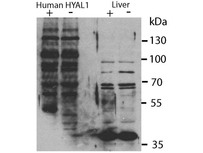
- Hyal1 +/+ mouse liver lysates, Hyal1 -/- mouse embryonic fibroblasts liver lysates overexpressing human HYAL1
- Negative Control
- Hyal1 -/- mouse liver lysates, Hyal1 -/- mouse embryonic fibroblasts
- Notes
- ABIN2784342 detects endogenous mouse HYAL1, and overexpressed human HYAL1 by Western blot analysis. The positive western blot assessment is based on the detection of a band of the appropriate molecular mass in Hyal1+/+ but not Hyal1-/- mouse tissue and the presence of a band of the appropriate molecular mass in Hyal1-/- cells transfected with human HYAL1. The identify of these bands was confirmed using the 1D10 antibody that had previously been shown to detect HYAL1. There are background bands to be considered in the interpretation of the blot.
- Primary Antibody
- ABIN2784342
- Secondary Antibody
- Peroxidase AffiniPure Donkey Anti-Rabbit IgG (H+L) (Jackson Labs, 711-035-152, Lot 106483)
- Full Protocol
- Overexpression of human HYAL1:
- The Human HYAL1 cDNA was cloned into an overexpressed vector and transiently transfected in mouse embryonic fibroblasts (MEFs) deficiency in HYAL1.
- Mouse Sample Preparation:
- Liver from Hyal1 knockout mice harvested along with that from a wildtype control was stored at -80°C until used.
- Liver pieces were weighed and placed in tubes containing 1.5ml of cold imidazole buffer pH7.4 containing 250mM sucrose with added protease inhibitors (Sigma).
- The tissues were homogenized by Polytron (Kinematica, PT10-35, two rounds of 5s at a setting of 6) with the tubes partially submerged in an ice bath to keep samples cold.
- The homogenized samples were centrifuged at 600xg for 10min to remove nuclei and intact cells.
- The post-nuclear supernatant was centrifuged at 100000xg for 1h.
- The resulting supernatant and resuspended pellet were measured in a Bradford assay for (Bio-Rad, 500-0006).
- Samples were boiled for 5min in SDS PAGE buffer including DTT as reducing reagent.
- Preliminary Western blot experiments with anti-Hyal1 (1D10) (hybridoma supernatant diluted 1:25) showed Hyal1 could specifically be detected in the 100000xg supernatant fraction of wildtype liver samples.
- This fraction was used for testing the other Hyal1 antibodies.
- Western blotting:
- Aliquots of the 100000xg supernatant fraction of wildtype liver sample corresponding to 30µg total protein were separated by SDS PAGE (7.5% gel) on Bio Rad II electrophoresis units.
- The separated proteins were transferred using Tris-glycine buffer (25mM tris, 192mM glycine pH8.3) for 1h at 100V to nitrocellulose membrane (Bio-Rad, 162-0112) using a Bio-Rad Trans-blot cell.
- Transfer quality was assessed by Ponceau S (0.1% in 3% acetic acid).
- Transfers were blocked with 5% non-fat milk in TBST (tris buffered saline + 0.1% Tween 20 pH7.4) for 1h at RT.
- Incubation with ABIN2784342 (1µg/ml) in 5ml in blocking buffer overnight at 4°C.
- Membranes were washed 3x 10min with TBST.
- Incubation with donkey-anti-rabbit secondary antibody conjugated to HRP (Jackson Labs, 711-035-152, Lot 106483) diluted 1:15000 in blocking buffer) for 1h at RT.
- Transfers were washed 3x 10min in TBST at RT.
- Transfers were washed 2x with H2O.
- Bands were visualized by incubation for 5min with Immobilon Western HRP Substrat (Millipore, WBKLS0500, lot 1530902).
- Image capture was carried on film (Kodak BioMax film for chemiluminescence) or digitally on a ChemiDoc (Bio-Rad).
- Experimental Notes
生效 #100030 (Western Blotting)
 Validation Images
![Western blot analysis of human HYAL1 overexpressed (first lane) in Hyal1 -/- MEFs (second lane). The two right lanes show detection enodgenous Hyal1 in mouse liver samples from Hyal1 +/+ (third lane) or Hyal1 -/- (fourth lane).]() Western blot analysis of human HYAL1 overexpressed (first lane) in Hyal1 -/- MEFs (second lane). The two right lanes show detection enodgenous Hyal1 in mouse liver samples from Hyal1 +/+ (third lane) or Hyal1 -/- (fourth lane).
Full Methods
Western blot analysis of human HYAL1 overexpressed (first lane) in Hyal1 -/- MEFs (second lane). The two right lanes show detection enodgenous Hyal1 in mouse liver samples from Hyal1 +/+ (third lane) or Hyal1 -/- (fourth lane).
Full Methods -
-
状态
- Liquid
-
浓度
- Lot specific
-
缓冲液
- Liquid. Purified antibody supplied in 1x PBS buffer with 0.09 % (w/v) sodium azide and 2 % sucrose.
-
储存液
- Sodium azide
-
注意事项
- This product contains Sodium azide: a POISONOUS AND HAZARDOUS SUBSTANCE which should be handled by trained staff only.
-
注意事项
- Avoid repeated freeze-thaw cycles.
-
储存条件
- -20 °C
-
储存方法
- For short term use, store at 2-8°C up to 1 week. For long term storage, store at -20°C in small aliquots to prevent freeze-thaw cycles.
-
-
- HYAL1 (Hyaluronidase-1 (HYAL1))
-
别名
- HYAL1
-
背景
-
HYAL1 is a lysosomal hyaluronidase. Hyaluronidases intracellularly degrade hyaluronan, one of the major glycosaminoglycans of the extracellular matrix. Hyaluronan is thought to be involved in cell proliferation, migration and differentiation. This enzyme is active at an acidic pH and is the major hyaluronidase in plasma. Mutations in HYAL1 are associated with mucopolysaccharidosis type IX, or hyaluronidase deficiency. HYAL1 is one of several related genes in a region of chromosome 3p21.3 associated with tumor suppression. Multiple transcript variants encoding different isoforms have been found for HYAL1.This gene encodes a lysosomal hyaluronidase. Hyaluronidases intracellularly degrade hyaluronan, one of the major glycosaminoglycans of the extracellular matrix. Hyaluronan is thought to be involved in cell proliferation, migration and differentiation. This enzyme is active at an acidic pH and is the major hyaluronidase in plasma. Mutations in this gene are associated with mucopolysaccharidosis type IX, or hyaluronidase deficiency. The gene is one of several related genes in a region of chromosome 3p21.3 associated with tumor suppression. Multiple transcript variants encoding different isoforms have been found for this gene.
Alias Symbols: HYAL-1, LUCA1, MGC45987, NAT6
Protein Interaction Partner: UBC, COL2A1,
Protein Size: 405 -
分子量
- 45 kDa
-
基因ID
- 3373
-
NCBI登录号
- NM_153282, NP_695014
-
UniProt
- Q12794
-
途径
- Glycosaminoglycan Metabolic Process
抗原
-


