C10orf2 抗体 (Middle Region)
 (2 references)
 (2 validations)
Our Local Distributor
北京 101111
Quick Overview for C10orf2 抗体 (Middle Region) (ABIN2775251)
抗原
See all C10orf2 (C10ORF2) 抗体适用
宿主
克隆类型
标记
应用范围
-
-
抗原表位
- Middle Region
-
原理
- TWNK Antibody - middle region
-
序列
- GVFRKFATDN NCHVTLVIHP RKEDDDKELQ TASIFGSAKA SQEADNVLIL
-
预测反应
- Cow: 100%, Dog: 100%, Guinea Pig: 100%, Horse: 100%, Human: 100%, Mouse: 100%, Rabbit: 100%, Rat: 100%, Zebrafish: 93%
-
产品特性
- This is a rabbit polyclonal antibody against PEO1. It was validated on Western Blot using a cell lysate as a positive control.
-
纯化方法
- Affinity Purified
-
免疫原
- The immunogen is a synthetic peptide directed towards the middle region of human PEO1
-
-
anti-Chromosome 10 Open Reading Frame 2 (C10ORF2) (AA 559-684) antibody
C10ORF2 适用: 人 WB, ELISA, IHC 宿主: 兔 Polyclonal unconjugated
anti-Chromosome 10 Open Reading Frame 2 (C10ORF2) (AA 656-684), (C-Term) antibodyC10ORF2 适用: 人 WB, IHC (p) 宿主: 兔 Polyclonal RB32016 unconjugated
anti-Chromosome 10 Open Reading Frame 2 (C10ORF2) (AA 385-684) antibodyC10ORF2 适用: 人 WB 宿主: 兔 Polyclonal unconjugated
anti-Chromosome 10 Open Reading Frame 2 (C10ORF2) (C-Term) antibodyC10ORF2 适用: 人, 小鼠, 大鼠 WB, IHC 宿主: 兔 Polyclonal unconjugated
anti-Chromosome 10 Open Reading Frame 2 (C10ORF2) antibodyC10ORF2 适用: 小鼠 WB 宿主: 兔 Polyclonal unconjugated
anti-Chromosome 10 Open Reading Frame 2 (C10ORF2) (AA 612-661) antibodyC10ORF2 适用: 人, 小鼠, 大鼠 WB 宿主: 兔 Polyclonal unconjugated
anti-Chromosome 10 Open Reading Frame 2 (C10ORF2) (AA 540-589) antibodyC10ORF2 适用: 人, 小鼠, 大鼠, Cow, 豚鼠, 马, 犬, 兔, Bat, 猴 WB 宿主: 兔 Polyclonal unconjugated
anti-Chromosome 10 Open Reading Frame 2 (C10ORF2) (C-Term) antibodyC10ORF2 适用: 人 WB 宿主: 兔 Polyclonal unconjugated
anti-Chromosome 10 Open Reading Frame 2 (C10ORF2) (AA 591-684) antibodyC10ORF2 适用: 人 ELISA, WB 宿主: 小鼠 Monoclonal 1C5 unconjugated
anti-Chromosome 10 Open Reading Frame 2 (C10ORF2) (AA 663-684), (C-Term) antibodyC10ORF2 适用: 人 WB, IHC (p), EIA 宿主: 兔 Polyclonal unconjugated
-
-
应用备注
- Optimal working dilution should be determined by the investigator.
-
说明
-
We also sell a specific blocking peptide that can be used in combination with this antibody. You can find the blocking peptide under AAP36483-100UG
-
限制
- 仅限研究用
-
-
- by
- Mitochondrial Biology, University of Eastern Finland
- No.
- #100038
- 日期
- 2016.08.08
- 抗原
- PEO1 antibody - middle region
- Lot Number
- QC6322
- Method validated
- Western Blotting
- Positive Control
- HeLa cells, HeLa cells overexpressing PEO1
- Negative Control
- rho0 HeLa cells
- Notes
- Passed, regarding sensitivity and specificity. The antibody detects an antigen at the expected apparent molecular weight of its antigen.
- Primary Antibody
- ABIN2775251
- Secondary Antibody
- Goat-anti rabbit IgG (H+L), HRP-linked (Invitrogen, A-16104)
- Full Protocol
- Cell lysates were propared from HeLa cells, HeLa cells overexpressing PEO1, and rho0 HeLa cells.
- 75µg total protein or each sample were separated on a denaturing 8% SDS-PAGE gel (Laemmli 1970).
- Immunoblot onto supported nitrocellulose membrane (Protran, GE Healthcare No. 1060005, 0.2µm) (Towbin et al., 1979).
- Blocking of the membrane in 3% skim milk in TBST (50mM Tris-HCl, pH 7.4, 150mM NaCl, 0.1% Tween 20) for 1h at room temperature.
- Incubation with primary antibody ABIN2775251 diluted 1:1000 in 3% BSA in TBST at 4°C overnight.
- Washing in TBST for 3x 5min.
- Incubation with goat-anti rabbit IgG (H+L), HRP-linked (Invitrogen, A-16104) diluted 1:15000 in 3% BSA in TBST for 1h at room temperature.
- Washing in TBST for 5x 10min.
- Chemiluminescence detection with luminol solution (250µg/ml Na-Luminol, 0,01% H2O2, 11µg/ml p-hydroxy-coumaric acid in 100mM Tris pH 8.5) and image capture on Kodak ECL film.
- Experimental Notes
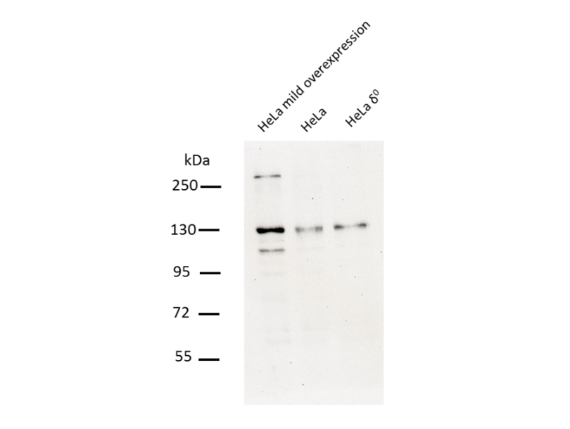
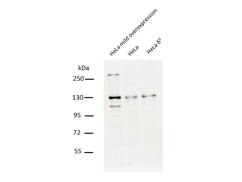
- PEO1 runs usually at 95-100 kDa, although the theoretical size is 72 kDa, often it is also visible as dimer.
生效 #100038 (Western Blotting)
 Validation Images
![75µg total protein of HeLa cells overexpressing PEO1 (lane 1), HeLa cells (lane 2), and rho0 Hela cells (lane 3) were separated on an denaturing 8% polyacrylamid gel. Bands were revealed as described in the protocol sections with ABIN2775251 diluted 1:1000.]() 75µg total protein of HeLa cells overexpressing PEO1 (lane 1), HeLa cells (lane 2), and rho0 Hela cells (lane 3) were separated on an denaturing 8% polyacrylamid gel. Bands were revealed as described in the protocol sections with ABIN2775251 diluted 1:1000.
Full Methods
75µg total protein of HeLa cells overexpressing PEO1 (lane 1), HeLa cells (lane 2), and rho0 Hela cells (lane 3) were separated on an denaturing 8% polyacrylamid gel. Bands were revealed as described in the protocol sections with ABIN2775251 diluted 1:1000.
Full Methods -
- by
- Mitochondrial Biology, University of Eastern Finland
- No.
- #100039
- 日期
- 2016.08.08
- 抗原
- PEO1 antibody - middle region
- Lot Number
- QC6322
- Method validated
- Immunocytochemistry
- Positive Control
- Mouse-anti-ds DNA antibody (clone 35I9)
- Negative Control
- Notes
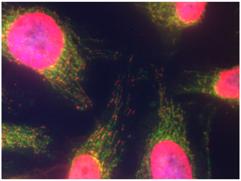
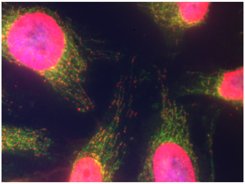
- Passed, regarding sensitivity and specificity. The antibody shows a staining pattern consistent with the expected mitochondrial localization of PEO1.
- Primary Antibody
- ABIN2775251
- Secondary Antibody
- Goat-anti-Rabbit IgG (H+L) Secondary Antibody, Alexa Fluor 488 conjugate (Invitrogen, A-11034, Lot 702323)
- Full Protocol
- HeLa cells were grown in DMEM high glucose + 10% FBS on acid-cleaned glass coverslips to 50% confluency.
- Cells were fixed in 4% para-formaldehyde in growth medium for 25min at RT, then washed once with growth medium and twice with PBS.
- Permeabilization was achieved by incubation with 0.5% Triton X-100, 10% fetal bovine serum in PBS for 15min.
- Primary antibodies:
- ABIN2775251 diluted 1:200 in PBS with 0.1% Triton X-100 , 10% fetal bovine serum.
- Co-staining of mitochondrial DNA with monoclonal antibody mouse-anti-dsDNA clone 35I9 DNA (Abcam ab27156, Lot GR27035-10) diluted 1:400 in PBS with 0.1% Triton X-100, 10% fetal bovine serum.
- Incubation for 1h at RT.
- Wash 3x 5min with PBS.
- Secondary antibodies:
- Goat anti-Rabbit IgG (H+L) Secondary Antibody, Alexa Fluor 488 conjugate (Invitrogen, A-11034, Lot 702323) diluted 1:1000 in PBS with 0,1% Triton X-100 , 10% fetal bovine serum, 1µM DAPI.
- Goat anti-Mouse IgG (H+L) Secondary Antibody, Alexa Fluor 594 conjugate (Invitrogen, A-11032, Lot 621333).
- Incubation for 1h at RT.
- Wash 3x 5min with PBS.
- The coverslips were mounted with Immu-mount mounting medium (Thermo Fisher, 9990402) and imaged with a 630 x magnification on a Zeiss Axioplan 2 fluorescent microscope using an Axiocam camera and Axiovision 4.8 software.
- For visualization of PEO1 the excitation wavelength was 488nm and emission wavelength >535nm, exposure time 53ms. DNA was visualized by excitation at 580nm and emission at >590nm, exposure time 410ms.
- Experimental Notes
生效 #100039 (Immunocytochemistry)
 Validation Images
![ABIN2775251 was used at a 1:200 dilution to visualize PEO1 (green, secondary antibody AF488 conjugate). A dsDNA antibody (red, secondary antibody AF594 conjugate) and a DAPI counterstain (blue) were used as reference.]() ABIN2775251 was used at a 1:200 dilution to visualize PEO1 (green, secondary antibody AF488 conjugate). A dsDNA antibody (red, secondary antibody AF594 conjugate) and a DAPI counterstain (blue) were used as reference.
Full Methods
ABIN2775251 was used at a 1:200 dilution to visualize PEO1 (green, secondary antibody AF488 conjugate). A dsDNA antibody (red, secondary antibody AF594 conjugate) and a DAPI counterstain (blue) were used as reference.
Full Methods -
-
状态
- Liquid
-
浓度
- 0.5 mg/mL
-
缓冲液
- Purified antibody supplied in 1x PBS buffer with 0.09 % (w/v) sodium azide and 2 % sucrose.
-
储存液
- Sodium azide
-
注意事项
- This product contains Sodium azide: a POISONOUS AND HAZARDOUS SUBSTANCE which should be handled by trained staff only.
-
注意事项
- prevent freeze-thaw cycles
-
储存条件
- 4 °C,-20 °C
-
储存方法
- For short term use, store at 2-8C up to 1 week. For long term storage, store at -20 °C in small aliquots to prevent freeze-thaw cycles.
-
-
-
: "Impaired p32 regulation caused by the lymphoma-prone RECQ4 mutation drives mitochondrial dysfunction." in: Cell reports, Vol. 7, Issue 3, pp. 848-58, (2014) (PubMed).
: "High mitochondrial DNA copy number has detrimental effects in mice." in: Human molecular genetics, Vol. 19, Issue 13, pp. 2695-705, (2010) (PubMed).
-
-
- C10orf2 (C10ORF2) (Chromosome 10 Open Reading Frame 2 (C10ORF2))
-
别名
- TWNK
-
背景
-
Background Information: This gene encodes a hexameric DNA helicase which unwinds short stretches of double-stranded DNA in the 5' to 3' direction and, along with mitochondrial single-stranded DNA binding protein and mtDNA polymerase gamma, is thought to play a key role in mtDNA replication. The protein localizes to the mitochondrial matrix and mitochondrial nucleoids. Mutations in this gene cause infantile onset spinocerebellar ataxia (IOSCA) and progressive external ophthalmoplegia (PEO) and are also associated with several mitochondrial depletion syndromes. Alternative splicing results in multiple transcript variants encoding distinct isoforms.
Gene Name: twinkle mtDNA helicase
Alternative Symbols: PEO, PEO1, SCA8, ATXN8, IOSCA, PEOA3, SANDO, TWINL, MTDPS7, PRLTS5, C10orf2
Protein Name: twinkle protein, mitochondrial
-
分子量
- 77 kDa
-
基因ID
- 56652
-
NCBI登录号
- NP_068602
-
UniProt
- Q96RR1
抗原
-