c-MYC 抗体 (AA 31-80)
 (2 validations)
Our Local Distributor
北京 101111
Quick Overview for c-MYC 抗体 (AA 31-80) (ABIN1532205)
抗原
See all c-MYC (MYC) 抗体适用
宿主
克隆类型
标记
应用范围
-
-
抗原表位
- AA 31-80
-
特异性
-
MYC Antibody detects endogenous levels of total MYC protein.
-
纯化方法
- The antibody was purified from rabbit antiserum by affinity-chromatography using immunogen.
-
纯度
- > 95 %
-
免疫原
- The antiserum was produced against synthesized peptide derived from human MYC.
-
亚型
- IgG
-
-
anti-Myc Proto-Oncogene protein (MYC) (AA 330-439) antibody
MYC 适用: 人 WB, IF, ELISA, IHC (p) 宿主: 小鼠 Monoclonal 1G7 unconjugated
anti-Myc Proto-Oncogene protein (MYC) (Center) antibodyMYC 适用: 人, 小鼠 WB, IF, IP, ICC, ChIP 宿主: 兔 Polyclonal unconjugated
anti-Myc Proto-Oncogene protein (MYC) (pSer62) antibodyMYC 适用: 人, 小鼠, 大鼠 WB, IHC, IF, ELISA, ICC 宿主: 兔 Polyclonal unconjugated
anti-Myc Proto-Oncogene protein (MYC) (pThr58) antibodyMYC 适用: 人, 小鼠, 大鼠 WB, IHC, IF, ELISA, IP, ICC 宿主: 兔 Polyclonal unconjugated
anti-Myc Proto-Oncogene protein (MYC) (pSer62) antibodyMYC 适用: 人, 小鼠, 大鼠 IHC, IF 宿主: 兔 Polyclonal unconjugated
anti-Myc Proto-Oncogene protein (MYC) (AA 360-440) antibodyMYC 适用: 人, 小鼠, 大鼠 WB, IF, ELISA, IHC (p) 宿主: 兔 Polyclonal unconjugated
anti-Myc Proto-Oncogene protein (MYC) (AA 101-200) antibodyMYC 适用: 人, 小鼠, 大鼠 WB, ELISA, IHC (p), ICC, IF (cc), IF (p), IHC (fro) 宿主: 兔 Polyclonal unconjugated
anti-Myc Proto-Oncogene protein (MYC) (N-Term) antibodyMYC 适用: 人, 小鼠, 大鼠 WB, IHC, IF, ELISA, ICC 宿主: 兔 Polyclonal unconjugated
anti-Myc Proto-Oncogene protein (MYC) antibodyMYC 适用: 人 ELISA 宿主: 小鼠 Monoclonal 7E10 unconjugated
anti-Myc Proto-Oncogene protein (MYC) (pThr58) antibodyMYC 适用: 人 WB, FACS, ICC 宿主: 兔 Monoclonal 24GB230 unconjugated Recombinant Antibody
-
-
应用备注
- WB: 1:500~1:1000 IHC: 1:50~1:100 IF: 1:100~1:500 ELISA: 1:40000
-
说明
-
Unigene-Number: Hs.202453 (NCBI Gene Symbol: MYC)
-
限制
- 仅限研究用
-
-
- by
- Developmental Biology, Johann-Friedrich-Blumenbach-Institute for Zoology and Anthropology, Georg-August-University of Göttingen
- No.
- #102753
- 日期
- 2018.07.03
- 抗原
- MYC
- Lot Number
- 210020 and 424170020
- Method validated
- Western Blotting
- Positive Control
HEK-293 cells transfected with Myc expression plasmid
- Negative Control
HEK-293 cells transfected with HA-Spag4-myc expression plasmid
- Notes
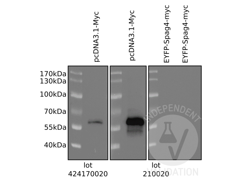
Passed. ABIN1532205 detects Myc protein in a western blot. No cross-reaction was observed with the myc-tag sequence positioned at the C-terminus of a recombinant protein.
- Primary Antibody
- ABIN1532205
- Secondary Antibody
- rabbit anti-mouse IgG, HRP-linked (Sigma A9044, Lot 034M4761)
- Full Protocol
- Grow HEK-293 cells (ATCC, CRL-1658) in DMEM+GlutaMAXDMEM+GlutaMAX (Gibco, 10567-014, lot 1922818) supplemented with fetal bovine serum (Gibco, 10500-064, lot 08FO477K) and Pen/Strep (Gibco, 15140-122, lot 1924797) at 37°C and 5% CO2 to 70% confluency.
- Transfect cells either with a Myc expression plasmid (pcDNA3.1-Myc) or HA- and myc-tagged SPAG4 expression plasmids using EndofectinMax (GeneCopoeia) following the manufacturer´s instructions.
- Grow cells for 24h.
- Lyse cells in SDS-sample buffer and denature total cellular lysates for 5min at 95°C. Subsequently separate them on a denaturing 10% SDS-PAGE (Laemmli 1970).
- Transfer proteins onto 0.2µm Protran membrane (GE Healthcare, 10600004, A10043108) by wet blotting for 1h at 400mA (Towbin et al., 1979).
- Block the membrane in TBST (50mM Tris-HCl, pH7.4, 150mM NaCl, 0.2% Tween 20) containing 5% milk (blocking solution) for 60min at RT.
- Incubate membrane with primary rabbit anti-myc antibody (antibodies-online, ABIN1532205 Lot#424170020 and Lot#210020) diluted 1:500 in blocking solution overnight at 4°C.
- Wash membrane with TBST for 45min at RT.
- Incubate membrane with secondary goat anti-rabbit IgG (H+L), HRP-linked (Jackson Immuno Research, 111-035-003, lot 123450) diluted 1:1000 in blocking solution for 45min at RT.
- Wash membrane 6x for 5min with TBST.
- Reveal protein bands using Clarity Max Western ECL substrate (Bio-Rad, 1705062) and capture images via Chemidoc Imaging System (BioRad).
- Incubate membrane with primary mouse anti-HA-tag antibody (clone 12CA5) diluted 1:500 in blocking solution overnight at 4°C.
- Wash membrane with TBST for 45min at RT.
- Incubate membrane with secondary rabbit anti-mouse IgG, HRP-linked (Sigma A9044, Lot 034M4761) diluted 1:1000 in blocking solution for 45min at RT.
- Reveal protein bands using Clarity Max Western ECL substrate (Bio-Rad, 1705062).
- Experimental Notes
In lysates of HEK-293 cells ectopically expressing recombinant protein with an N-terminal HA- and a C-terminal myc-tag ABIN1532205 did not detect the labeled protein at the expected molecular weight. In contrast, an anti-HA-tag antibody (clone 12CA5) revealed a protein at the expected MW.
生效 #102753 (Western Blotting)
 Validation Images
![ABIN1532205 lot 424170020 (let panel) and lot 210020 (middle and right panel) detected a protein in the expected molecular mass range exclusively when Myc is expressed (pcDNA3.1-Myc) but did not react with the Myc-tagged SPAG4 (EYFP-Spag4-myc; expected MW approximately 50kDa).]() ABIN1532205 lot 424170020 (let panel) and lot 210020 (middle and right panel) detected a protein in the expected molecular mass range exclusively when Myc is expressed (pcDNA3.1-Myc) but did not react with the Myc-tagged SPAG4 (EYFP-Spag4-myc; expected MW approximately 50kDa).
Full Methods
ABIN1532205 lot 424170020 (let panel) and lot 210020 (middle and right panel) detected a protein in the expected molecular mass range exclusively when Myc is expressed (pcDNA3.1-Myc) but did not react with the Myc-tagged SPAG4 (EYFP-Spag4-myc; expected MW approximately 50kDa).
Full Methods -
- by
- Developmental Biology, Johann-Friedrich-Blumenbach-Institute for Zoology and Anthropology, Georg-August-University of Göttingen
- No.
- #103023
- 日期
- 2018.07.03
- 抗原
- MYC
- Lot Number
- 210020 and 424170020
- Method validated
- Immunocytochemistry
- Positive Control
HEK-293 cells transfected with Myc expression plasmid
- Negative Control
HEK-293 cells transfected with HA-Spag4-myc expression plasmid
- Notes
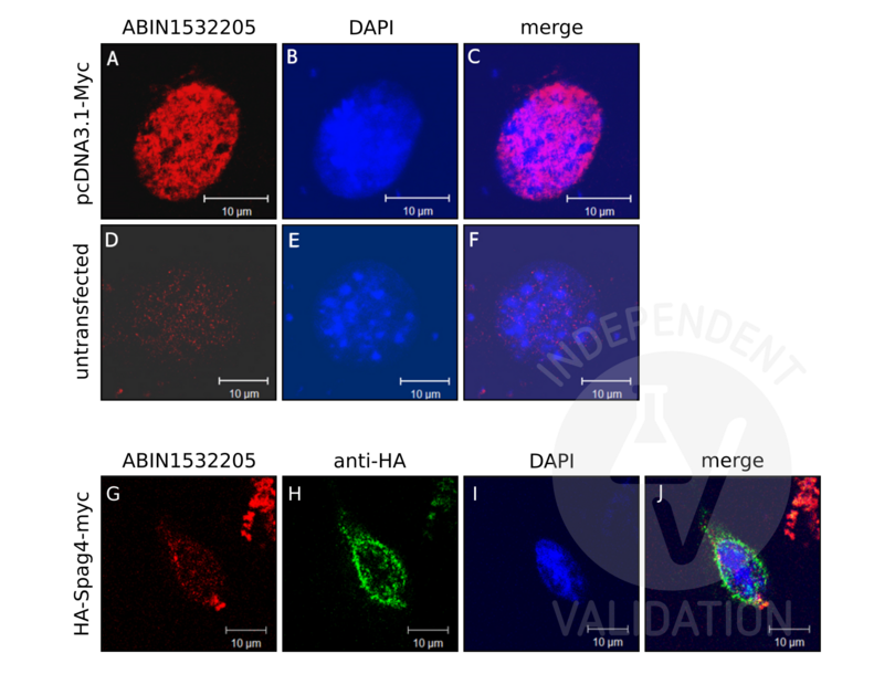
Passed. ABIN1532205 (lot 424170020 and lot 210020) detects Myc in immunocytochemistry. No cross-reaction was observed with the myc-tag sequence positioned at the C-terminus of a recombinant protein.
- Primary Antibody
- ABIN1532205
- Secondary Antibody
- goat anti-rabbit IgG MFP590 (Mobitec MFP-A1037, lot 3002088), F(ab')2-Goat anti-Rabbit IgG (H+L) Alexa Fluor 555 (Life Technologies, A21430, lot 1637266)
- Full Protocol
- Grow HEK-293 cells (ATCC, CRL-1658) in DMEM+GlutaMAXDMEM+GlutaMAX (Gibco, 10567-014, lot 1922818) supplemented with fetal bovine serum (Gibco, 10500-064, lot 08FO477K) and Pen/Strep (Gibco, 15140-122, lot 1924797) at 37°C and 5% CO2 to 70% confluency.
- Transfect cells either with a Myc expression plasmid (pcDNA3.1-Myc) or HA- and myc-tagged SPAG4 expression plasmids using EndofectinMax (GeneCopoeia) following the manufacturer´s instructions.
- Grow cells for 24h.
- Fix cells in 3.7% paraformaldehyde (in PBS) for 20min at 4°C followed by incubation in 0.3% Triton X-100 for 10min at 4°C.
- Block cells in PBS containing 1% BSA and 0.5% Tween-20 (PBT) for 1h at RT.
- Incubate cells with primary antibody
- rabbit anti-Myc antibody (antibodies-online, ABIN1532205, lot 424170020 or lot 210020) diluted 1:50 in PBS and mouse anti-HA tag antibody (clone 12CA5) ON at 4°C (images G, H).
- rabbit anti-Myc antibody (antibodies-online, ABIN1532205, lot 210020 or lot 210020) diluted 1:50 in PBS ON at 4°C (images A, D).
- Wash cells with 50mM Tris-HCl, pH7.4, 150mM NaCl, 0.1% Tween 20 (TBST) for 15min at RT.
- Incubate cells with secondary antibody
- goat anti-rabbit IgG MFP590 (Mobitec MFP-A1037, lot 3002088) diluted 1:300 in PBS and goat anti-mouse IgGDylight488 (Thermo Scientific, 35503) diluted 1:10000 in PBS for 1h at 37°C (images G, H).
- F(ab')2-Goat anti-Rabbit IgG (H+L) Alexa Fluor 555 (Life Technologies, A21430, lot 1637266) diluted 1:2000 in PBS for 45min at 37°C (images A, D).
- Counterstain cells with DAPI (Sigma, D-9542).
- Image acquisition on Zeiss LSM 510 confocal microscope and processing using Adobe Photoshop 5.0.
- Experimental Notes
The antibody detects Myc proteins by immunoblotting and by immunocytology. No cross-reaction with the C-terminal Myc-tag.
生效 #103023 (Immunocytochemistry)
 Validation Images
![ABIN1532205 detects ectopically expressed Myc (pcDNA3.1-Myc) (A, red). No cross-reactivity is observed in untransfected cells (D). DAPI was used to visualize the nucleus (B and E, blue). Images on the right show the merged red and blue channels. ABIN1532205 did not detect the myc-tag (G, red) of HA-Spag4-myc whereas an anti HA-tag antibody (H, green) detected HA-Spag4-myc at the nuclear membrane as expected. The picture on the right (J) shows the red and green channels merged with DAPI counterstain (I, blue).]() ABIN1532205 detects ectopically expressed Myc (pcDNA3.1-Myc) (A, red). No cross-reactivity is observed in untransfected cells (D). DAPI was used to visualize the nucleus (B and E, blue). Images on the right show the merged red and blue channels. ABIN1532205 did not detect the myc-tag (G, red) of HA-Spag4-myc whereas an anti HA-tag antibody (H, green) detected HA-Spag4-myc at the nuclear membrane as expected. The picture on the right (J) shows the red and green channels merged with DAPI counterstain (I, blue).
Full Methods
ABIN1532205 detects ectopically expressed Myc (pcDNA3.1-Myc) (A, red). No cross-reactivity is observed in untransfected cells (D). DAPI was used to visualize the nucleus (B and E, blue). Images on the right show the merged red and blue channels. ABIN1532205 did not detect the myc-tag (G, red) of HA-Spag4-myc whereas an anti HA-tag antibody (H, green) detected HA-Spag4-myc at the nuclear membrane as expected. The picture on the right (J) shows the red and green channels merged with DAPI counterstain (I, blue).
Full Methods -
-
状态
- Liquid
-
浓度
- 1 mg/mL
-
缓冲液
- phosphate buffered saline (without Mg2+ and Ca2+), pH 7.4, 150 mM NaCl, 0.02 % sodium azide and 50 % glycerol.
-
储存液
- Sodium azide
-
注意事项
- This product contains sodium azide: a POISONOUS AND HAZARDOUS SUBSTANCE which should be handled by trained staff only.
-
储存条件
- -20 °C
-
储存方法
- Stable at -20°C for at least 1 year.
-
有效期
- 12 months
-
-
- c-MYC (MYC) (Myc Proto-Oncogene protein (MYC))
-
别名
- Myc Proto-Oncogene protein
-
背景
-
Synonyms: Myc proto-oncogene protein, Class E basic helix-loop-helix protein 39, bHLHe39, Proto-oncogene c-Myc, Transcription factor p64 , MYC , BHLHE39
NCBI Gene Symbol: MYC
-
分子量
- 48 kDa
-
基因ID
- 4609
-
OMIM
- 113970
-
UniProt
- P01106
-
途径
- p53 Pathway, Cell Division Cycle, Sensory Perception of Sound, Transition Metal Ion Homeostasis, Mitotic G1-G1/S Phases, Positive Regulation of Endopeptidase Activity, Regulation of Carbohydrate Metabolic Process, Positive Regulation of Response to DNA Damage Stimulus, Warburg Effect
抗原
-


