C19ORF80 ELISA 试剂盒


 (10 references)
 (1 validation)
Our Local Distributor
北京 101111
Quick Overview for C19ORF80 ELISA 试剂盒 (ABIN1136417)
抗原
See all C19ORF80 ELISA试剂盒适用
检测方法
实验类型
检测范围
应用范围
样品类型
-
-
最低检测浓度
- 78 pg/mL
-
Analytical Method
- Quantitative
-
灵敏度
- 54pg/ml
-
产品特性
- Homo sapiens,Human,Betatrophin,Angiopoietin-like protein 8,Lipasin,C19orf80,Angptl8,UNQ599/PRO1185
-
组件
-
Reagent (Quantity):
- Assay plate (1),
- Standard (2),
- Sample Diluent (1×20 mL),
- Assay Diluent A (1×10 mL),
- Assay Diluent B (1×10 mL),
- Detection Reagent A (1×120 μL),
- Detection Reagent B (1×120 μL),
- Wash Buffer(25 x concentrate) (1×30 mL),
- Substrate (1×10 mL),
- 2 Stop Solution (1×10 mL),
- Plate sealer for 96 wells (5),
- Instruction (1)
-
试剂未包括
- Microplate reader. Pipettes and pipette tips. EP tube Deionized or distilled water.
-
-
Chromosome 19 Open Reading Frame 80 (C19ORF80) ELISA Kit
C19ORF80 适用: 小鼠 Colorimetric Sandwich ELISA 15.6 pg/mL - 1000 pg/mL Cell Culture Supernatant, Cell Lysate, Plasma, Serum, Tissue Homogenate
Chromosome 19 Open Reading Frame 80 (C19ORF80) ELISA KitC19ORF80 适用: 大鼠 Colorimetric Sandwich ELISA 31.2 pg/mL - 2000 pg/mL Plasma, Serum, Tissue Homogenate
Chromosome 19 Open Reading Frame 80 (C19ORF80) ELISA KitSmall Sample C19ORF80 适用: 人 Colorimetric Sandwich ELISA 0.156 ng/mL - 10 ng/mL Cell Culture Supernatant, Cell Lysate, Plasma, Serum, Tissue Homogenate
Chromosome 19 Open Reading Frame 80 (C19ORF80) ELISA KitC19ORF80 适用: 人 Colorimetric Sandwich ELISA 46.875 pg/mL - 3000 pg/mL Plasma, Serum, Tissue Homogenate
Chromosome 19 Open Reading Frame 80 (C19ORF80) ELISA KitC19ORF80 适用: 小鼠 Colorimetric Sandwich ELISA 0.313 ng/mL - 20 ng/mL Plasma, Serum, Tissue Homogenate
Chromosome 19 Open Reading Frame 80 (C19ORF80) ELISA KitC19ORF80 适用: 人 Colorimetric 6.25-400 pg/mL Plasma, Serum, Tissue Homogenate
Chromosome 19 Open Reading Frame 80 (C19ORF80) ELISA KitC19ORF80 适用: 大鼠 Colorimetric Sandwich ELISA 78.12 pg/mL - 5000 pg/mL Cell Culture Supernatant, Cell Lysate, Plasma, Serum, Tissue Homogenate
-
-
说明
-
Gene Name: C19orf80
-
样本量
- 100 μL
-
板类型
- Pre-coated
-
实验流程
- The microtiter plate provided in this kit has been pre-coated with an antibody specific to the target. Standards or samples are then added to the appropriate microtiter plate wells with a biotin-conjugated polyclonal antibody preparation specific for target and Avidin conjugated to Horseradish Peroxidase (HRP) is added to each microplate well and incubated. Then a TMB (3,3'5, 5' tetramethyl-benzidine) substrate solution is added to each well. Only those wells that contain the target, biotin-conjugated antibody and enzyme-conjugated Avidin will exhibit a change in color. The enzyme-substrate reaction is terminated by the addition of a sulphuric acid solution and the color change is measured spectrophotometrically at a wavelength of 450 nm ± 2 nm. The concentration of target in the samples is then determined by comparing the O.D. of the samples to the standard curve.
-
试剂准备
-
Bring all reagents to room temperature before use. Wash Buffer - If crystals have formed in the concentrate, warm to room temperature and mix gently until the crystals have completely dissolved. Dilute 30 mL of Wash Buffer Concentrate into deionized or distilled water to prepare 750 mL of Wash Buffer. Standard - Reconstitute the Standard with 1.0 mL of Sample Diluent. This reconstitution produces a stock solution. Allow the standard to sit for a minimum of 15 minutes with gentle agitation prior to making serial dilutions (Making serial dilution in the wells directly is not permitted). The undiluted standard serves as the high standard. The Sample Diluent serves as the zero standard (0 ng/ml).
-
样品收集
-
Serum - Use a serum separator tube (SST) and allow samples to clot for 30 minutes before centrifugation for 15 minutes at approximately 1000 × g. Remove serum and assay immediately or aliquot and store samples at -20 °C or -80 °C.
Plasma - Collect plasma using EDTA or heparin as an anticoagulant. Centrifuge samples for 15 minutes at 1000 × g at 2 °C - 8 °C within 30 minutes of collection. Store samples at -20 °C or -80 °C. Avoid repeated freeze-thaw cycles.
Tissue homogenates - The preparation of tissue homogenates will vary depending upon tissue type. For this assay, tissue was rinsed with 1X PBS to remove excess blood, homogenized in 20 mL of 1X PBS and stored overnight at ≤ -20 °C After two freeze-thaw cycles were performed to break the cell membranes, the homogenates were centrifuged for 5 minutes at 5000 x g. Remove the supernate and assay immediately or aliquot and store at ≤ -20 °C.
Cell culture supernates and Other biological fluids - Remove particulates by centrifugation and assay immediately or aliquot and store samples at -20 °C or -80 °C. Avoid repeated freeze-thaw cycles.
Note:
1. Samples to be used within 5 days may be stored at 2-8 °C , otherwise samples must stored at -20 °C (1 month) or -80 °C (2 months) to avoid loss of bioactivity and contamination.
2. Tissue or cell extraction samples prepared by chemical lysis buffer may cause unexpected ELISA results due to the impacts of certain chemicals.
3. Influenced by the factors including cell viability, cell number and also sampling time, samples from cell culture supernatant may not be detected by the kit
4. Sample hemolysis will influence the result, so hemolytic specimen can not be detected.
5. When performing the assay slowly bring samples to room temperature. -
实验流程
-
Allow all reagents to reach room temperature (Please do not dissolve the reagents at 37 °C directly.). All the reagents should be mixed thoroughly by gently swirling before pipetting. Avoid foaming. Keep appropriate numbers of strips for 1 experiment and remove extra strips from microtiter plate. Removed strips should be resealed and stored at 4 °C until the kits expiry date. Prepare all reagents, working standards and samples as directed in the previous sections. Please predict the concentration before assaying. If values for these are not within the range of the standard curve, users must determine the optimal sample dilutions for their particular experiments.
1. Add 100 μL of Standard, Blank, or Sample per well. Cover with the Plate sealer. Incubate for 2 hours at 37 °C .
2. Remove the liquid of each well, don ’ t wash.
3. Add 100 μL of Detection Reagent A working solution to each well. Cover with the Plate sealer. Incubate for 1 hour at 37 °C . Detection Reagent A working solution may appear cloudy. Warm to room temperature and mix gently until solution appears uniform.
4. Aspirate each well and wash, repeating the process three times for a total of three washes. Wash by filling each well with Wash Buffer (approximately 400 μL) using a squirt bottle, multi-channel pipette, manifold dispenser or autowasher. Complete removal of liquid at each step is essential to good performance. After the last wash, remove any remaining Wash Buffer by aspirating or decanting. Invert the plate and blot it against clean paper towels.
5. Add 100 μL of Detection Reagent B working solution to each well. Cover with a new Plate sealer. Incubate for 1 hours at 37 °C .
6. Repeat the aspiration/wash as in step 4.
7. Add 90 μL of Substrate Solution to each well. Cover with a new Plate sealer. Incubate within 30 minutes at 37 °C . Protect from light.
8. Add 50 μL of Stop Solution to each well. If color change does not appear uniform, gently tap the plate to ensure thorough mixing.
9. Determine the optical density of each well at once, using a microplate reader set to 450 nm.
Important Note:
1. Absorbance is a function of the incubation time. Therefore, prior to starting the assay it is recommended that all reagents should be freshly prepared prior to use and all required strip-wells are secured in the microtiter frame. This will ensure equal elapsed time for each pipetting step, without interruption.
2. Please carefully reconstitute Standards or working Detection Reagent A and B according to the instruction, and avoid foaming and mix gently until the crystals have completely dissolved. The reconstituted Standards can be used only once. This assay requires pipetting of small volumes. To minimize imprecision caused by pipetting, ensure that pipettors are calibrated. It is recommended to suck more than 10 μ l for once pipetting.
3. To ensure accurate results, proper adhesion of plate sealers during incubation steps is necessary. Do not allow wells to sit uncovered for extended periods between incubation steps. Once reagents have been added to the well strips, DO NOT let the 5 strips DRY at any time during the assay.
4. For each step in the procedure, total dispensing time for addition of reagents to the assay plate should not exceed 10 minutes.
5. To avoid cross-contamination, change pipette tips between additions of each standard level, between sample additions, and between reagent additions. Also, use separate reservoirs for each reagent.
6. The wash procedure is critical. Insufficient washing will result in poor precision and falsely elevated absorbance readings.
7. Duplication of all standards and specimens, although not required, is recommended.
8. Substrate Solution is easily contaminated. Please protect it from light. -
结果分析
-
Average the duplicate readings for each standard, control, and sample and subtract the average zero standard optical density. Create a standard curve by reducing the data using computer software capable of generating a four parameter logistic (4-PL) curve-fit. As an alternative, construct a standard curve by plotting the mean absorbance for each standard on the x-axis against the concentration on the y-axis and draw a best fit curve through the points on the graph. The data may be linearized by plotting the log of the SAA concentrations versus the log of the O.D. and the best fit line can be determined by regression analysis. It is recommended to use some related software to do this calculation, such as curve expert 13.0. This procedure will produce an adequate but less precise fit of the data. If samples have been diluted, the concentration read from the standard curve must be multiplied by the dilution factor.
-
限制
- 仅限研究用
-
-
- by
- Shakti Bioresearch
- No.
- #029581
- 日期
- 2014.01.26
- 抗原
- Lot Number
- 3L306L
- Method validated
- ELISA
- Positive Control
- Human serum
- Negative Control
- Mouse serum
- Notes
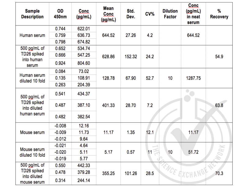
- Matrix interference indicates that serum must be diluted >10 fold for accurate measurement. Kit returned minor signal for negative control sample.
- Primary Antibody
- Antigen: Human Chromosome 19 Open Reading Frame 80 (C19ORF80)
- Catalog number: E11644h
- Lot number: 3L306L
- Secondary Antibody
- Full Protocol
- All reagents in the ELISA kit were brought up to room temperature (RT) before use.
- 100 µl of each sample was added per well to the micro ELISA plate well. All samples and standards were assayed in triplicate.
- The plate was covered with sealer (provided in kit) and incubated for 120 mins at 37°C.
- Liquid was removed from each well by pipette.
- Detection Reagent A was diluted 100 fold in Assay Diluent A. 100 µl of diluted detection reagent A was added to each well and the plate was sealed. The plate was tapped to ensure mixing and incubated for 60 mins at 37°C.
- Wells were washed with 300 µl wash buffer three times. Each wash involved fully aspirating the liquid from each well by pipette. After the last wash the plate was inverted against clean absorbent paper to remove any remaining liquid.
- Detection Reagent B was diluted 100 fold in Assay Diluent B. 100 µl of diluted detection reagent B was added to each well and the plate was sealed. The plate was tapped to ensure mixing and incubated for 60 mins at 37°C.
- Wells were washed with 300 µl wash buffer five times. Each wash involved fully aspirating the liquid from each well by pipette. After the last wash the plate was inverted against clean absorbent paper to remove any remaining liquid.
- 90 µl of Substrate Solution was added to each well and the plate was covered with a new plate sealer. The plate was tapped to ensure mixing and incubated at room temperature in the dark.
- After about 10 mins, when an apparent gradient appeared in the standard wells, the reaction was terminated by adding 50 µl of Stop Solution to each well.
- The optical density (OD value) of each well was read using a micro-plate reader set to 450 nm.
- The triplicate readings for each sample were averaged and the average zero standard optical density subtracted. A standard curve was generated by plotting the mean OD value for each standard on the y-axis against the concentration on the x-axis using Softmax Pro softare.
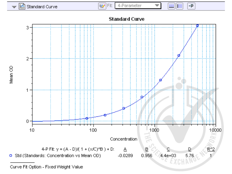
- The equation y = (A-D)/(1 + (x/C)^B) + D was used to calculate IL-6 concentrations of the samples based on their average OD values.
- Experimental Notes
- Percent recovery of the spiked samples shows that there is matrix interference. Dilution of >10 fold is required for accurate measurement of the analyte in human serum samples.
生效 #029581 (ELISA)
 Validation Images
![Figure 1: Graph of corrected-average absorbance (OD 450 nm) readings plotted for standard curve samples.]() Figure 1: Graph of corrected-average absorbance (OD 450 nm) readings plotted for standard curve samples.
Figure 1: Graph of corrected-average absorbance (OD 450 nm) readings plotted for standard curve samples.
![Table 1: ELISA. C19ORF80 could be detected in human serum (positive control). Spike controls indicate that there is interference from the human serum matrix and a dilution of >10 fold is required. Mouse serum was used as negative control, there were residual levels of C19ORF80.]() Table 1: ELISA. C19ORF80 could be detected in human serum (positive control). Spike controls indicate that there is interference from the human serum matrix and a dilution of >10 fold is required. Mouse serum was used as negative control, there were residual levels of C19ORF80.
Table 1: ELISA. C19ORF80 could be detected in human serum (positive control). Spike controls indicate that there is interference from the human serum matrix and a dilution of >10 fold is required. Mouse serum was used as negative control, there were residual levels of C19ORF80.
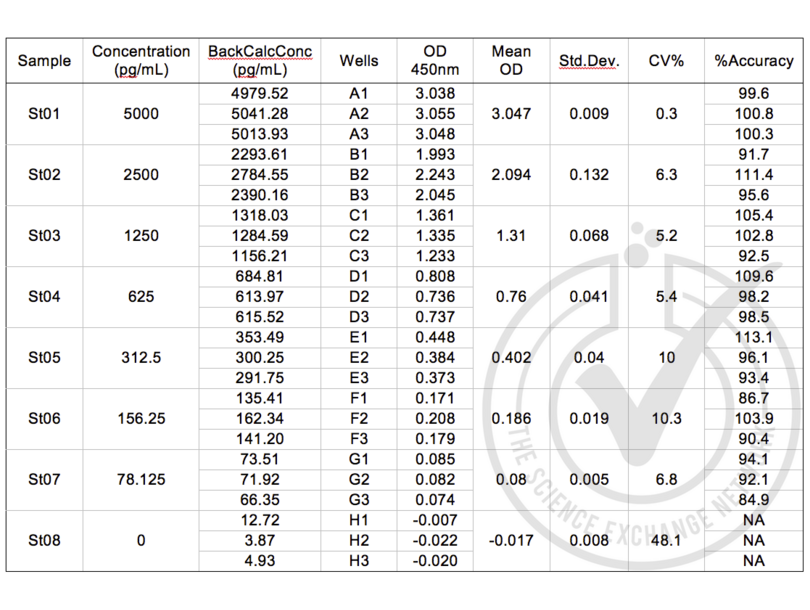
![Table 2: Table of absorbance readings (OD 450 nm) for standard curve. Value for Average Reading is derived from the average of three readings (OD 450nm). The Average Reading for BLANK (0 pg/ml) was subtracted from all Average Readings to yield Average Absorbance values for Standards. Standard deviation is included for all samples. An equation (see Figure 1) was generated from the standard curve and used to calculate C19ORF80 concentrations shown in Table 1.]() Table 2: Table of absorbance readings (OD 450 nm) for standard curve. Value for Average Reading is derived from the average of three readings (OD 450nm). The Average Reading for BLANK (0 pg/ml) was subtracted from all Average Readings to yield Average Absorbance values for Standards. Standard deviation is included for all samples. An equation (see Figure 1) was generated from the standard curve and used to calculate C19ORF80 concentrations shown in Table 1.
Full Methods
Table 2: Table of absorbance readings (OD 450 nm) for standard curve. Value for Average Reading is derived from the average of three readings (OD 450nm). The Average Reading for BLANK (0 pg/ml) was subtracted from all Average Readings to yield Average Absorbance values for Standards. Standard deviation is included for all samples. An equation (see Figure 1) was generated from the standard curve and used to calculate C19ORF80 concentrations shown in Table 1.
Full Methods -
-
储存条件
- 4 °C/-20 °C
-
储存方法
- The Assay Plate, Standard, Detection Reagent A and Detection Reagent B should be stored at -20°C upon being received. After receiving the kit , Substrate should be always stored at 4°C.
-
-
-
: "Correlation of circulating betatrophin concentrations with insulin secretion capacity, evaluated by glucagon stimulation tests." in: Diabetic medicine : a journal of the British Diabetic Association, Vol. 32, Issue 5, pp. 653-6, (2016) (PubMed).
: "Increased levels of irisin in people with long-standing Type 1 diabetes." in: Diabetic medicine : a journal of the British Diabetic Association, Vol. 32, Issue 9, pp. 1172-6, (2016) (PubMed).
: "Relationship of betatrophin with youth onset type 2 diabetes among Asian Indians." in: Diabetes research and clinical practice, Vol. 109, Issue 1, pp. 71-6, (2016) (PubMed).
: "Characterisation of betatrophin concentrations in childhood and adolescent obesity and insulin resistance." in: Pediatric diabetes, Vol. 17, Issue 1, pp. 53-60, (2016) (PubMed).
: "Associations of betatrophin levels with irisin in Chinese women with normal glucose tolerance." in: Diabetology & metabolic syndrome, Vol. 7, pp. 26, (2015) (PubMed).
: "Increased circulating levels of betatrophin in newly diagnosed type 2 diabetic patients." in: Diabetes Care, Vol. 37, Issue 10, pp. 2718-22, (2014) (PubMed).
: "Elevated circulating lipasin/betatrophin in human type 2 diabetes and obesity." in: Scientific reports, Vol. 4, pp. 5013, (2014) (PubMed).
: "An explanation for recent discrepancies in levels of human circulating betatrophin." in: Diabetologia, Vol. 57, Issue 10, pp. 2232-4, (2014) (PubMed).
: "Circulating betatrophin correlates with atherogenic lipid profiles but not with glucose and insulin levels in insulin-resistant individuals." in: Diabetologia, Vol. 57, Issue 6, pp. 1204-8, (2014) (PubMed).
: "Increased circulating betatrophin concentrations in patients with type 2 diabetes." in: International journal of endocrinology, Vol. 2014, pp. 323407, (2014) (PubMed).
-
-
- C19ORF80 (Chromosome 19 Open Reading Frame 80 (C19ORF80))
-
别名
- C19orf80
-
背景
- Synonyms: C19orf80,Hepatocellular carcinoma-associated protein TD26,Homo sapiens,Human,UNQ599/PRO1185
抗原 See all C19ORF80 ELISA试剂盒
-