RDH10 抗体
 (1 reference)
 (1 validation)
Our Local Distributor
北京 101111
Quick Overview for RDH10 抗体 (ABIN7118460)
抗原
See all RDH10 抗体适用
宿主
克隆类型
标记
应用范围
-
-
原理
- RDH10 antibody
-
纯化方法
- Immunogen affinity purified
-
纯度
- ≥95 % as determined by SDS-PAGE
-
免疫原
- retinol dehydrogenase 10(all-trans)
-
亚型
- IgG
-
-
anti-Retinol Dehydrogenase 10 (All-Trans) (RDH10) (AA 106-135) antibody
RDH10 适用: 人 WB, FACS, IHC (p) 宿主: 兔 Polyclonal RB25251 unconjugated
anti-Retinol Dehydrogenase 10 (All-Trans) (RDH10) (Internal Region) antibodyRDH10 适用: 人, 大鼠, 小鼠 WB, IHC, ICC, IF 宿主: 兔 Polyclonal unconjugated
anti-Retinol Dehydrogenase 10 (All-Trans) (RDH10) (AA 71-120) antibodyRDH10 适用: 人, 大鼠, 小鼠, Cow, 犬, 马, 兔, Bat, 猴, Pig WB, IHC, IHC (p) 宿主: 兔 Polyclonal unconjugated
anti-Retinol Dehydrogenase 10 (All-Trans) (RDH10) (AA 242-291) antibodyRDH10 适用: 人, 大鼠, 小鼠, Cow, 犬, 马, 兔, 豚鼠, 斑马鱼, Bat, 猴, Pig, 非洲爪蟾 WB 宿主: 兔 Polyclonal unconjugated
anti-Retinol Dehydrogenase 10 (All-Trans) (RDH10) (AA 1-341) antibodyRDH10 适用: 人, 大鼠 WB, ELISA, FACS 宿主: 兔 Polyclonal unconjugated
-
-
应用备注
- WB: 1:500-1:2000, IP: 1:200-1:1000, IHC: 1:20-1:200
-
限制
- 仅限研究用
-
-
- by
- Palczewski Lab, Center For Translational Vision Research, UC Irvine
- No.
- #104469
- 日期
- 2023.03.23
- 抗原
- RDH10
- Lot Number
- 20221223E
- Method validated
- Immunohistochemistry
- Positive Control
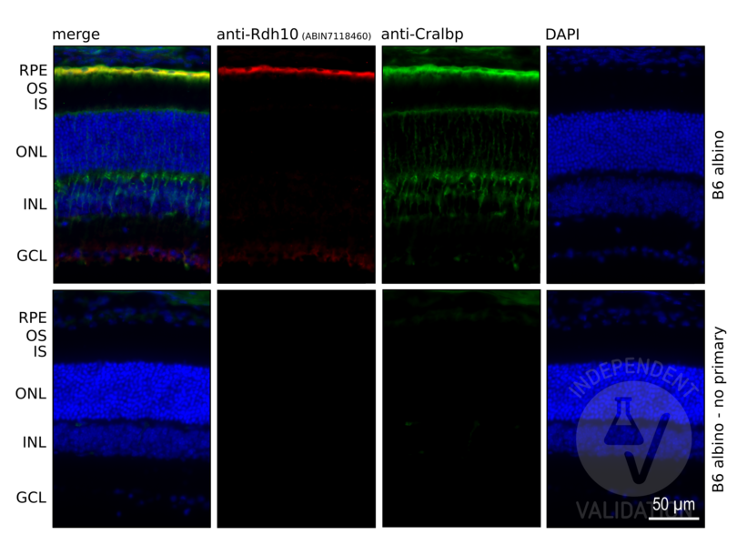
Retina cryosection from B6 Albino (B6(Cg)-Tyrc-2J/J) animal
- Negative Control
Retina cryosection from B6 Albino (B6(Cg)-Tyrc-2J/J) animal
No primary antibody
- Notes
Passed. Presence of specific signal in the RPE cell layer was considered as indication of specific immunoreactivity using the anti-RDH10 antibody ABIN7118460.
- Primary Antibody
- ABIN7118460
- Secondary Antibody
- donkey anti-rabbit AF647-conjugated antibody (Abcam, 150075)
- Full Protocol
- Collect eyes from mice and fix with paraformaldehyde 4% (Electron Microscopy Sciences, 15710) in 1x PBS for 30 min at RT.
- Cryoprotection with sucrose series:
- Wash in 10% sucrose in 1x PBS.
- Immerse in 10% sucrose in 1x PBS for 30 min at RT.
- Wash in 20% sucrose in 1x PBS.
- Immerse in 20% sucrose in 1x PBS for 30 min RT.
- Wash in 30% sucrose in 1x PBS.
- 30% sucrose ON at 4°C.
- Embed eyes in OCT compound (Tissue-Tek O.C.T. Compound, 4583).
- Cut retinal sections at a thickness of 12 μm on a cryostat.
- Air dry sections for 15 min at RT, store at -80°C until use.
- Bring sections to RT and rehydrate in 1x PBS for 1 h.
- Incubate sections in blocking buffer (1x PBS, 3% BSA (Sigma-Aldrich, A7030), 3% Donkey serum (Sigma-Aldrich, S30-100ML), 0.1% Triton X-100 (Sigma-Aldrich, X100-500ML)) for 1 h at RT.
- Incubate sections with primary rabbit anti-RDH10 antibody (antibodies-online, ABIN7118460, lot 20221223E) diluted 1:50 in blocking buffer ON at RT. Include a no primary antibody negative controls. Additionally, counterstaing with primary mouse anti-CRALBP antibody (Thermo Fisher Scientific, MA1-813).
- Rinse sections 3 times with 1x PBS, 0.1% Triton X100. Keep negative controls in a separate container.
- Incubate sections with secondary AF647-conjugated donkey anti-rabbit antibody (Abcam, Ab150075) or AF488-conjugated donkey anti-mouse antibody (Thermo Fisher Scientific, A32766) diluted 1:500 in blocking buffer for 1 h at RT.
- Rinse sections once with 1x PBS, 0.1% Triton X-100 for 5 min at RT.
- Incubate sections in 1x DAPI (Thermo Fisher Scientific, 62248) in 1x PBS, 0.1% Triton X-100 for 15 min at RT.
- Rinse sections 3x with 1x PBS, 0.1% Triton X-100 for 5 min at RT.
- Mount sections in VECTASHIELD® HardSet™ Antifade Mounting Medium (Vector Laboratories, H-1400) mounting medium.
- Acquire images with a fluorescence microscope and appropriate filter settings.For the validation purposes Keyence BZ-X800E fluorescence microscope was used with following filters: BZ-X DAPI for DAPI, BZ-X GFP for AF488, BZ-X Cy5 for AF647. Images were taken at 10x and 40x magnification.
- Experimental Notes
Experiment involved validation of the specificity of 4 antibodies against different Rdh proteins: Rdh5 (ABIN7254060), Rdh10 (ABIN7118460), Rdh11 (ABIN966957), and Rdh12 (ABIN7167836). All 4 proteins are important for eye function and highly expressed in neural retina and/or RPE. Validation is based on comparison of each staining with known pattern of expression in the mouse retina. For review highlighting each Rdh localization see PMID20801113.
To aid orientation in the cell layers anti-Cralbp counterstain was included in the staining (Thermo MA1-813). Cralbp (Rlbp1) is highly expressed in RPE and Müller glia cells in mouse retina.
生效 #104469 (Immunohistochemistry)
 Validation Images
![Retinal sections from the wild-type (B6 albino) mice immunostained with anti-Rdh10 antibody ABIN7118460. DAPI staining shows localization of the inner (INL) and outer (ONL) nuclear layer of the mouse retina. Cralbp (Rlbp1) co-staining was used to visualize RPE and Müller glia cells in the retina. Presence of specific signal in the RPE cell layer confirms specific immunoreactivity.]() Retinal sections from the wild-type (B6 albino) mice immunostained with anti-Rdh10 antibody ABIN7118460. DAPI staining shows localization of the inner (INL) and outer (ONL) nuclear layer of the mouse retina. Cralbp (Rlbp1) co-staining was used to visualize RPE and Müller glia cells in the retina. Presence of specific signal in the RPE cell layer confirms specific immunoreactivity.
Full Methods
Retinal sections from the wild-type (B6 albino) mice immunostained with anti-Rdh10 antibody ABIN7118460. DAPI staining shows localization of the inner (INL) and outer (ONL) nuclear layer of the mouse retina. Cralbp (Rlbp1) co-staining was used to visualize RPE and Müller glia cells in the retina. Presence of specific signal in the RPE cell layer confirms specific immunoreactivity.
Full Methods -
-
状态
- Liquid
-
缓冲液
- PBS with 0.02 % sodium azide and 50 % glycerol pH 7.3,
-
储存液
- Sodium azide
-
注意事项
- This product contains Sodium azide: a POISONOUS AND HAZARDOUS SUBSTANCE which should be handled by trained staff only.
-
注意事项
- Avoid repeated freeze / thaw cycles.
-
储存条件
- -20 °C
-
储存方法
- -20°C for 12 months
-
有效期
- 12 months
-
-
-
: "Rapid RGR-dependent visual pigment recycling is mediated by the RPE and specialized Müller glia." in: Cell reports, Vol. 42, Issue 8, pp. 112982, (2023) (PubMed).
-
-
- RDH10 (Retinol Dehydrogenase 10 (All-Trans) (RDH10))
-
别名
- RDH10
-
背景
-
Synonyms: Retinol dehydrogenase 10|Short chain dehydrogenase/reductase family 16C member 4|RDH10|SDR16C4
Background: Retinol dehydrogenase with a clear preference for NADP. Converts all-trans-retinol to all-trans-retinal. Has no detectable activity towards 11-cis-retinol, 9-cis-retinol and 13-cis-retinol.
-
分子量
- 39 kDa
-
基因ID
- 157506
-
UniProt
- Q8IZV5
抗原
-


