Biotin 抗体 (DyLight 800)
 (1 validation)
Our Local Distributor
北京 101111
Quick Overview for Biotin 抗体 (DyLight 800) (ABIN6720668)
抗原
宿主
克隆类型
标记
应用范围
-
-
Supplier Product No.
- 600-445-098
-
Supplier
- Rockland
-
原理
- Biotin Antibody Dylight™ 800 Conjugated
-
交叉反应 (详细)
- Assay by immunoelectrophoresis resulted in a single precipitin arc against anti-Rabbit Serum, Biotin conjugated IgG and Biotin conjugated Bovine Serum Albumin.
-
纯化方法
- This product was prepared from monospecific antiserum by immunoaffinity chromatography using Biotin coupled to sepharose beads.
-
免疫原
- Biotin conjugated to Keyhole Limpet Hemocyanin (KLH)
-
亚型
- IgG
-
Labeling Ratio
- 2.5
-
-
anti-Biotin antibody
WB, IHC, IP, ICC 宿主: 兔 Monoclonal BLR315M unconjugated Recombinant Antibody
anti-Biotin antibodyWB, IHC, IP, ICC 宿主: 兔 Monoclonal BLR315M unconjugated Recombinant Antibody
anti-Biotin antibodyWB, IHC, ICC 宿主: 兔 Monoclonal BLR328M unconjugated Recombinant Antibody
anti-Biotin antibody适用: Various Species IHC 宿主: 兔 Monoclonal BTN-2032R unconjugated Recombinant Antibody
anti-Biotin antibody适用: Various Species WB, IHC, FACS, IF 宿主: 小鼠 Monoclonal Hyb-8 unconjugated
anti-Biotin antibodyWB, IHC, FACS, ELISA, IF, FM 宿主: 小鼠 Monoclonal 4C7-D5 unconjugated
anti-Biotin antibody适用: 人 WB 宿主: 兔 Monoclonal unconjugated
anti-Biotin antibodyFACS, ELISA, IP, MS 宿主: 小鼠 Monoclonal 1D4-C5 unconjugated
-
-
应用备注
-
FLISA_Dilution: >1:20,000
IF_Microscopy_Dilution: >1:5,000
Western_Blot_Dilution: 1:10,000 - 1:25,000
Other: User Optimized
-
说明
-
Anti-Biotin DyLight 800 Conjugated Antibody has been tested by western blot. This product is designed for immunofluorescence microscopy, fluorescence based plate assays (FLISA) and fluorescent western blotting. This product is also suitable for multiplex analysis, including multicolor imaging, utilizing various commercial platforms. The emission spectra for this DyLight™ conjugate match the principle output wavelengths of most common fluorescence instrumentation.
-
限制
- 仅限研究用
-
-
- by
- Prof. Merighi, Laboratory of Neurobiology, Department of Veterinary Sciences, University of Turin
- No.
- #104645
- 日期
- 2026.11.09
- 抗原
- Biotin
- Lot Number
- 24716
- Method validated
- Immunohistochemistry
- Positive Control
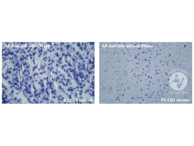
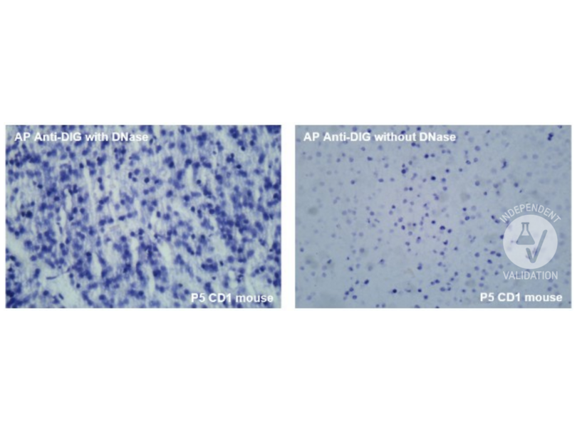
Sections from the same tissues were incubated with an anti-digoxigenin antibody conjugated with alkaline phosphatase (AP) and developed with NBT/BCIP solution.
- Negative Control
Omission of Terminal transferase.
- Notes
The antibody works in IHC-P at a concentration of 1:500-1:5000.
- Primary Antibody
- ABIN6720668
- Secondary Antibody
- Full Protocol
- Sample analyzed: P5 CD1 mouse brain
- Paraffin-embedded slices (7 µm thick) were deparaffinized and rehydrated through a graded series of alcohols and distilled water (dH2O).
- Sections were incubated in proteinase K [20 µg/mL] for 15 minutes at room temperature (RT).
- 4 × 2 minutes washing in double distilled water (ddH2O).
- Sections were briefly washed in DN buffer and incubated in DNase [1 µg/mL] for 10 minutes at RT.
- 4 × 2 minutes washing in ddH2O.
- 5 minutes washing in TDT buffer at RT.
- Sections were incubated in TDT buffer added with Terminal transferase [0.05 U/µL] and digoxigenin-11-dUTP [10 µM] for 2 hours at +37 °C.
- 15 minutes washing in TB buffer at RT.
- 5 minutes washing in ddH2O + 5 minutes washing in 0.01 M PBS.
- Sections were incubated with the primary antibody at the following dilutions of 1:50, 1:200, 1:500, overnight at +4 °C (primary antibody was diluted in PLL/BSA/PBS diluent added with 5% BSA).
- 3 × 5-minute washes in 0.01 M PBS.
- Incubation with the anti-mouse secondary antibody 1:1000 in PLL/BSA/PBS diluent, 1 hour at RT.
- 3 × 5-minute washes in 0.01 M PBS.
- Nuclear counterstaining was performed with 4′,6-Diamidine-2′-phenylindole dihydrochloride (DAPI) [1 µg/mL], 2 minutes.
- 2 × 5 minutes washing in 0.01 M PBS.
- Specimens were mounted in Fluoroshield mounting medium (Sigma).
- Images were acquired with a Leica DM 6000B fluorescence microscope with a digital camera at 40× magnification.
OTHER PERTINENT REAGENTS USED:
- Proteinase K (Thermo Scientific, Catalog # EO0491)
- DNase I (in the kit by Sigma, Catalog # AMP-D1)
- Terminal transferase (in the kit by Jena Biosciences, Catalog # APP-002)
- Digoxigenin-11-dUTP (Roche, Catalog # 11573179910)
- DN buffer: Tris base [30 mM] pH 7.2, sodium cacodylate [140 mM], magnesium chloride [4 mM], DTT [0.1 mM] in dH2O
- TDT buffer: Tris base [30 mM] pH 7.2, sodium cacodylate [140 mM], cobalt chloride [1 mM] in dH2O
- TB buffer: sodium chloride [300 mM], sodium citrate [30 mM] in dH2O
- Experimental Notes
To exclude any nonspecific signal due to the secondary antibody, one control slice was processed for each experimental procedure, omitting the primary antibody.
生效 #104645 (Immunohistochemistry)
 Validation Images
![Positive control with Alkaline Phosphatase Anti-Digoxigenin antibody on P5 CD1 mouse brain. Images show reaction with and without DNase treatment to increase cellular death. Magnification: 40x.]() Positive control with Alkaline Phosphatase Anti-Digoxigenin antibody on P5 CD1 mouse brain. Images show reaction with and without DNase treatment to increase cellular death. Magnification: 40x.
Positive control with Alkaline Phosphatase Anti-Digoxigenin antibody on P5 CD1 mouse brain. Images show reaction with and without DNase treatment to increase cellular death. Magnification: 40x.
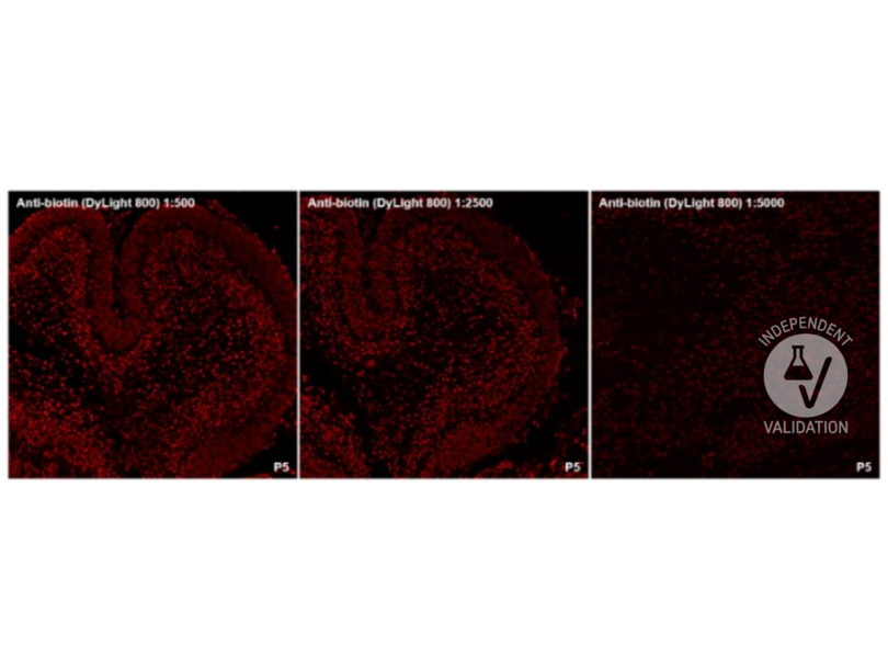
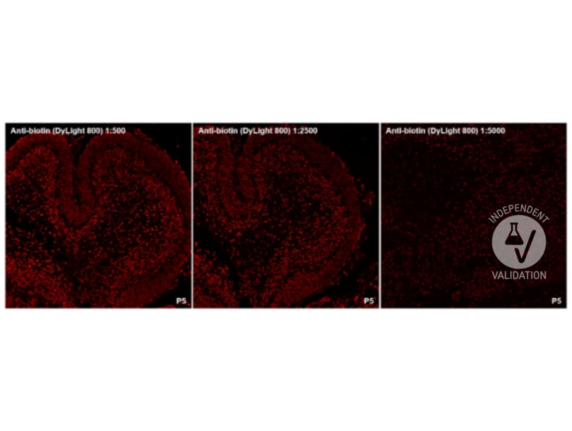
![The TUNEL reaction was performed using biotinylated dUTP and revealed with ABIN6720668 (anti-biotin DyLight 800-conjugated). Magnification: 20x.]() The TUNEL reaction was performed using biotinylated dUTP and revealed with ABIN6720668 (anti-biotin DyLight 800-conjugated). Magnification: 20x.
Full Methods
The TUNEL reaction was performed using biotinylated dUTP and revealed with ABIN6720668 (anti-biotin DyLight 800-conjugated). Magnification: 20x.
Full Methods -
-
状态
- Lyophilized
-
溶解方式
-
Reconstitution_Buffer: Restore with deionized water (or equivalent)
Reconstitution_Volume: 100 μL
-
缓冲液
-
Buffer: 0.02 M Potassium Phosphate, 0.15 M Sodium Chloride, pH 7.2
Stabilizer: 10 mg/mL Bovine Serum Albumin (BSA) - Immunoglobulin and Protease free
Preservative: 0.01 % (w/v) Sodium Azide -
储存液
- Sodium azide
-
注意事项
- This product contains Sodium azide: a POISONOUS AND HAZARDOUS SUBSTANCE which should be handled by trained staff only.
-
储存条件
- 4 °C,-20 °C
-
储存方法
- Store vial at 4° C prior to restoration. For extended storage aliquot contents and freeze at -20° C or below. Avoid cycles of freezing and thawing. Centrifuge product if not completely clear after standing at room temperature. This product is stable for several weeks at 4° C as an undiluted liquid. Dilute only prior to immediate use.
-
有效期
- 12 months
-
-
- Biotin
-
物质类
- Chemical
-
背景
-
Synonyms: rabbit anti-Biotin Antibody Dylight™ 800 Conjugated, rabbit anti Biotin Antibody Dylight™ 800 Conjugated
Background: Biotin Antibody detects Biotin. Biotin is a water-soluble B-complex vitamin (vitamin B7). It is composed of a ureido (tetrahydroimidizalone) ring fused with a tetrahydrothiophene ring. A valeric acid substituent is attached to one of the carbon atoms of the tetrahydrothiophene ring. Biotin is a coenzyme for carboxylase enzymes, involved in the synthesis of fatty acids, isoleucine, and valine, and in gluconeogenesis. Biotin is necessary for cell growth, the production of fatty acids, and the metabolism of fats and amino acids. Anti-Biotin DyLight Conjugated Antibody is ideal for investigators involved in Cell Signaling and Cell Biology research.
抗原
-